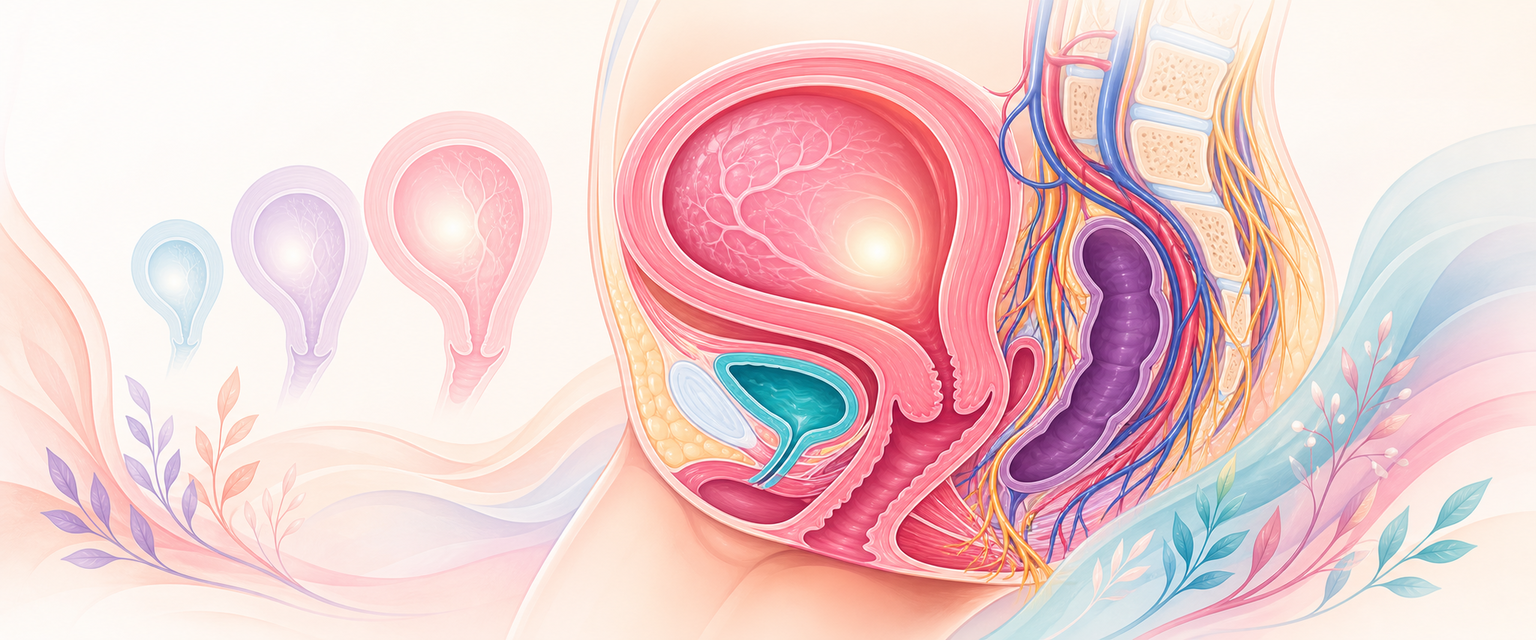
Pregnant Uterus
Start from the simplest fact and let everything else follow from it: the uterus is a hollow bag of smooth muscle whose job is to hold a pregnancy quietly for nine months and then expel it in a few hours. Every anatomical change in pregnancy serves one of those two opposite jobs — containment (grow large, stay quiet, keep blood flowing to the placenta) or delivery (contract powerfully, dilate the outlet, then clamp shut to stop bleeding). If you keep asking "is this serving containment or delivery?", the whole chapter assembles itself.
Pregnancy turns a small pelvic organ into a large, high-flow, contractile abdominal organ. The same transformation explains normal fetal growth, the mechanics of labour, where the surgeon cuts at caesarean, why a scar can rupture, why a placenta sits low, why a placenta can invade, and why the uterus bleeds catastrophically when it fails to contract. A candidate who understands the pregnant uterus reasons from first principles rather than memorising isolated complications.
The organising idea is this: pregnancy remodels the wall, cavity, cervix, placental bed, blood supply, supports, peritoneal relations and nerve pathways — but at different speeds and for different purposes. Most clinical problems arise when one part does not match the demand placed on it: a poorly formed lower segment, a scarred placental bed, an atonic myometrium, a distorted bladder reflection, a stretched scar or an overdistended uterus. This chapter stays at the level of anatomy and mechanism; recognition and treatment of the complications it explains are carried by the linked Intermediate and Final chapters.
Non-Pregnant Baseline
You cannot describe a change without first fixing the starting point, so anchor on the resting organ before pregnancy touches it.
The non-pregnant uterus has a fundus, body, isthmus and cervix. It lies between bladder and rectum, usually anteverted (tilted forward on the vagina) and anteflexed (bent forward at the isthmus), with the uterovesical pouch anteriorly and the pouch of Douglas posteriorly. Its wall has three broad layers: endometrium, myometrium and serosa/peritoneum.
| Part | Baseline anatomy | Pregnancy importance |
|---|---|---|
| Fundus and body | Thick muscular upper uterus | Main expanding and contractile segment |
| Isthmus | Narrow region between body and cervix | Major contributor to lower uterine segment |
| Cervix | Collagen-rich canal with glands | Maintains pregnancy, remodels and dilates |
| Endometrium | Cycling mucosa | Becomes decidua after implantation |
| Myometrium | Interlacing smooth muscle | Hypertrophies, stretches and contracts around vessels |
| Serosa/peritoneum | Covers fundus/body; reflections to bladder and rectum | Surgical planes change as uterus enlarges |
The non-pregnant uterine artery reaches the cervix after crossing above the ureter in the base of the broad ligament — "water under the bridge", the bridge being the artery and the water the ureter. This relationship is a permanent surgical rule. Pregnancy increases the size, flow and fragility of the vessels; it does not move the ureter out of danger during lateral extensions, hysterectomy or distorted repeat caesarean.
Two further baseline facts matter once the uterus enlarges. First, the uterus is held by supports that pregnancy does not create but does stretch: the transverse cervical (cardinal) ligaments that anchor the cervix laterally and carry the uterine vessels, the uterosacral ligaments that pull the cervix backwards and maintain anteversion, and the round ligaments that run from the uterine cornua through the inguinal canal to the labia majora. Second, the uterus has an orderly lymphatic drainage: the cervix and lower uterus drain to the obturator and internal/external iliac nodes, the upper body drains to the para-aortic nodes, and the cornual/round-ligament region drains to the superficial inguinal nodes. These supports and drainage routes are the same in pregnancy; what changes is the load and the blood flow passing through them.
First Trimester: Decidua, Implantation and a Pelvic Uterus
In the first trimester the uterus is still mainly pelvic. It softens, becomes more vascular and enlarges under hormonal influence. The endometrium becomes decidua, a specialised pregnancy mucosa that supports implantation, regulates trophoblast invasion and forms the maternal part of the placental interface.
| Change | Mechanism | Clinical bridge |
|---|---|---|
| Decidualisation | Progesterone-dominant stromal transformation | Implantation support, decidual bleeding |
| Trophoblast invasion | Extravillous trophoblast enters decidua and spiral arteries | Placental bed, pre-eclampsia and PAS concepts |
| Myometrial hypertrophy/hyperplasia | Smooth muscle cells enlarge and increase early | Uterine growth before major stretch |
| Cervical vascularity | Increased blood flow and gland activity | Bluish cervix, contact bleeding, mucus plug |
| Corpus luteum support | Progesterone production before placental dominance | Early pregnancy/adnexal relevance |
Decidualisation is worth singling out because it is the maternal half of the placental interface and the gatekeeper of trophoblast invasion. Progesterone — supplied first by the corpus luteum and, after the luteoplacental shift in the early second trimester, by the placenta itself — drives the endometrial stroma to become decidua. This same progesterone is the dominant containment signal that keeps the myometrium quiet (the thread picked up later under labour onset).
A uterus smaller than expected may reflect wrong dates, miscarriage, ectopic pregnancy or fetal growth problems later. A uterus larger than expected may reflect multiple pregnancy, fibroids or trophoblastic disease. Those clinical differentials are learned elsewhere; the anatomy here is that early uterine size and softness are still assessed against a pelvic organ with palpable cervix, body and adnexa. With the decidua now in place, the next step is what the trophoblast does to the maternal vessels beneath it — the placental bed.
Placental Bed and Spiral Artery Transformation
The placental bed is the decidua and underlying myometrium beneath the placenta. It is not just a surface attachment. It contains maternal decidual cells, immune cells, glands, myometrial fibres, spiral arteries and extravillous trophoblast.
Normal placentation requires controlled trophoblast invasion. Extravillous trophoblast has two broad components:
| Component | Location/action | Result |
|---|---|---|
| Interstitial trophoblast | Moves through decidua and superficial myometrium | Anchors placenta and remodels tissue interface |
| Endovascular trophoblast | Enters spiral arteries | Replaces/changes vessel wall to create low-resistance flow |
In the non-pregnant state, spiral arteries are muscular, reactive vessels designed for cyclical endometrial shedding — they constrict to limit menstrual blood loss. In pregnancy they must do the opposite: become dilated, flaccid, low-resistance, funnel-shaped uteroplacental channels that deliver a large, steady, pressure-buffered stream of maternal blood into the intervillous space. Trophoblast achieves this by replacing the endothelium and destroying the muscular and elastic media of the vessel wall, so the artery can no longer narrow.
Remodelling happens in two waves. The first wave transforms the decidual (superficial) segments of the spiral arteries early in pregnancy. A second, deeper wave extends the change into the myometrial segments through roughly the second trimester, with adequate transformation normally complete by about 24 weeks. The endpoint is continuous, low-resistance maternal flow with little vasoreactivity — the placental bed becomes, in effect, a high-volume vascular sink. This deep two-wave conversion is what differentiates a healthy placental bed from one that will hypoperfuse the fetus.
Mechanism-to-clinical-consequence chain:
| Mechanism | Anatomical effect | Clinical association |
|---|---|---|
| Inadequate spiral artery remodelling | Persistently narrow, reactive vessels | Placental hypoperfusion, fetal growth restriction, pre-eclampsia risk |
| Placenta implants low in developing lower segment | Placental edge near internal os | Placenta praevia/low-lying placenta concepts |
| Decidual deficiency over scar | Trophoblast approaches or invades myometrium | Placenta accreta spectrum |
| Placental separation from normal decidual plane | Exposed uteroplacental vessels compressed by myometrium | Normal third stage haemostasis |
| No normal cleavage plane in PAS | Placenta remains attached/invades | Major haemorrhage risk if disrupted |