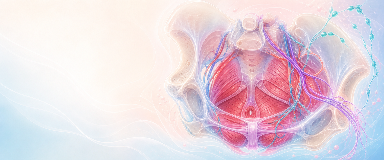
Female Perineum
The perineum is the outlet of the pelvis. It is where the bony pelvis, pelvic floor, anal sphincter complex, distal vagina, urethra, vulva, erectile tissues, vessels, nerves and lymphatics meet in a small space. In O&G it is also where birth stretch, episiotomy, obstetric tears, vulval haematoma, pudendal block, dyspareunia, continence and later prolapse become clinically visible. The internal pudendal artery is one branch of the pelvic arterial tree set out in Pelvic anatomy and supports.
The single most useful idea for the whole chapter is this: the perineum is not just skin between the vagina and anus. It is a layered fibromuscular support system with its own predictable map of muscles, fascia, sphincters, vessels and nerves. When a force passes through it during birth or surgery, the clinical consequence depends entirely on which layer fails — mucosa, superficial muscles, perineal body, anal sphincter, anorectal mucosa, pudendal branches, erectile tissue, venous plexus or levator support. Everything that follows is an attempt to make that map automatic, so that in theatre you can name the structure your finger or needle is on before you act.
We will build the chapter from the outside in and from simple to complex: first the surface boundaries you can draw, then the two triangles, then the layered compartments, then the perineal body that ties them together, then continence anatomy, then the spaces that hide blood, then the single neurovascular bundle that supplies almost all of it, then lymphatics, and finally how all of this behaves under the stretch of birth.
Surface Map and Boundaries
Start with what you can see and draw. The anatomical perineum is the diamond-shaped region inferior to the pelvic diaphragm (the levator ani and coccygeus that close the pelvic floor from above). Its boundaries are fixed bony and ligamentous points, so the same drawing works in an exam and the same orientation works in theatre.
| Boundary | Structure |
|---|---|
| Anterior point | Pubic symphysis |
| Anterolateral borders | Ischiopubic rami |
| Lateral points | Ischial tuberosities |
| Posterolateral borders | Sacrotuberous ligaments |
| Posterior point | Coccyx |
A transverse line joining the two ischial tuberosities (which passes just anterior to the anus) divides the diamond into an anterior urogenital triangle and a posterior anal triangle. Memorise this line: it is the reference for almost every other structure in the perineum, and it is the line an episiotomy and most obstetric tears cross.
| Region | Main contents | O&G relevance |
|---|---|---|
| Urogenital triangle | Distal urethra, distal vagina, vestibule, perineal membrane, erectile tissue, superficial perineal muscles, Bartholin glands | Episiotomy, second-degree tears, vulval haematoma, dyspareunia, urethral support |
| Anal triangle | Anal canal, internal and external anal sphincters, ischioanal fossae, pudendal canal branches | OASIS, continence, perianal sepsis, pudendal block |
| Perineal body | Midline fibromuscular node between distal vagina and anal canal | Posterior vaginal support, genital hiatus size, repair quality |
The clinically useful drawing starts with the perineal body as the anchor. Place it in the midline between the posterior vaginal wall and the anterior anal canal, then attach to it the bulbospongiosus, superficial transverse perineal muscles, external anal sphincter and fibres related to the levator ani and rectovaginal septum. That one point explains why an apparently local perineal tear can simultaneously affect posterior vaginal support, sexual pain and anal continence.
The Vulva and Vestibule
Before going deep, anchor the surface structures of the urogenital triangle, because these are what you actually retract, infiltrate and repair, and because their names recur throughout O&G.
The labia majora are the outer fatty folds covered by skin; they contain hair follicles, sebaceous glands and apocrine (modified sweat) glands. The labia minora are the smaller inner folds; they are devoid of adipose tissue and have no hair follicles, and they meet anteriorly to form the prepuce of the clitoris and posteriorly merge into the fourchette.
The vestibule is the area bounded by the clitoris anteriorly, the labia minora laterally, the fourchette posteriorly and the hymen superiorly. Two sets of glands open into it, and both matter clinically:
| Structure | Position | Clinical relevance |
|---|---|---|
| Skene (paraurethral) ducts | Either side of the external urethral meatus, anteriorly | Skene duct cyst/abscess, periurethral pathology |
| Bartholin (greater vestibular) glands | Open at 5 and 7 o'clock posterolaterally, just inside the introitus | Bartholin cyst/abscess — a very common gynaecology presentation |
The Bartholin glands are mucoid, alkaline-secreting glands arranged as lobules of alveoli lined by cuboidal or columnar epithelium; their ducts open into the posterolateral vestibule. The clitoris consists of two erectile corpora cavernosa that terminate in the sensitive glans; its crura attach to the medial aspects of the ischiopubic rami. The paired vestibular bulbs are masses of erectile tissue lying at the sides of the vestibule, deep to the labia, covered by bulbospongiosus. This erectile/venous tissue is exactly why a tear or episiotomy extension here can bleed briskly or form a tense haematoma even when the skin breach looks modest. The lower vagina, vestibule and vulval skin all sit in this single anterior territory, so a single injury can cross several of these named structures at once.
Layered Urogenital Triangle
Now go deep. The urogenital triangle is learned superficial to deep, because each layer has a different failure pattern during birth or surgery and a different repair priority.
| Layer | Contents | Clinical consequence when injured |
|---|---|---|
| Skin and superficial fascia | Vulval/perineal skin, subcutaneous fat, superficial veins | Bruising, superficial tears, vulval haematoma |
| Superficial perineal pouch | Bulbospongiosus, ischiocavernosus, superficial transverse perineal muscles, vestibular bulbs, Bartholin glands | Second-degree tears, episiotomy tissue plane, bleeding from erectile/venous tissue |
| Perineal membrane | Tough fascial sheet spanning the pubic arch | Distal urethral and vaginal support; barrier between superficial and deep compartments |
| Deep perineal pouch | External urethral sphincter, deep transverse perineal muscles, neurovascular branches, areolar tissue | Urethral continence and deep support |
| Pelvic diaphragm above | Levator ani and coccygeus | Pelvic organ support, hiatal size, levator trauma |
The perineal membrane is a dense fascial sheet that attaches to the sides of the urogenital triangle (the ischiopubic rami) and is pierced by the urethra and the vagina. It is not merely a passive partition: it contributes to the distal support of the urethra and vagina and provides an attachment platform for the perineal muscles. When the membrane and perineal body lose integrity, the genital hiatus widens and the levator hiatus is less well supported from below.