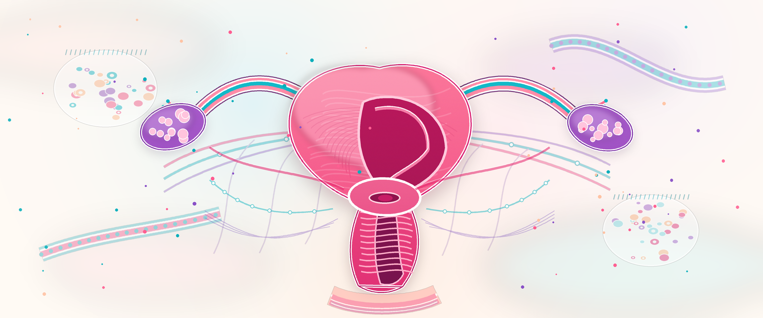
Female Genital Tract Anatomy and Histology
The female genital tract is a continuous system from vulval skin to ovarian cortex, but each region has its own surface, epithelium, stromal structure, vascular supply, nerve supply, lymph drainage and disease pattern. O&G uses this anatomy in every examination and operation: vulval inspection, speculum sampling, cervical screening, labour, caesarean section, hysterectomy, salpingectomy, oophorectomy, prolapse repair, endometriosis surgery, cancer staging and STI care.
The single first principle that makes the whole tract make sense is its embryological origin. The internal tract — uterine tubes, uterus, cervix and upper vagina — is built from the paired paramesonephric (Müllerian) ducts that fuse in the midline. The lower vagina and vestibule arise from the urogenital sinus, and the external vulva forms from the genital tubercle, folds and swellings. The ovary, by contrast, is a gonad that develops separately on the posterior abdominal wall and descends only as far as the pelvis. Three consequences flow directly from this and recur through the chapter: (1) the tube opens freely into the peritoneal cavity because the Müllerian duct never sealed at its cranial end; (2) the change from columnar Müllerian lining to squamous urogenital-sinus lining happens at the cervix, which is exactly why the cervix has a junction that becomes the screening target; and (3) the ovary keeps its high abdominal blood supply (ovarian artery off the aorta) and high lymphatic drainage (para-aortic nodes) despite sitting low in the pelvis, because its vessels followed it down from where it was born. Hold these three facts and most of the surgical and oncological anatomy is predictable rather than memorised.
The safest way to learn the tract is then outside-in and surface-to-deep:
- vulva: skin, mucocutaneous vestibule, glands, erectile tissue and pudendal territory;
- vagina: fibromuscular canal with non-keratinised squamous epithelium and no glands;
- cervix: ectocervix, endocervical canal and transformation zone;
- uterus: endometrium, myometrium, cervix, peritoneal reflections and support complex;
- uterine tubes: fimbriae, infundibulum, ampulla, isthmus and intramural part;
- ovaries: follicles, stroma, corpus luteum, surface covering, hilum and vascular pedicle.
| Region | Dominant histology | Key O&G meaning |
|---|---|---|
| Vulva | Keratinised skin plus mucocutaneous vestibule | Dermatoses, pain mapping, Bartholin disease, vulval cancer nodes |
| Vagina | Non-keratinised stratified squamous epithelium | Discharge, atrophy, prolapse, repair planes |
| Ectocervix | Stratified squamous epithelium | Visible cervix, squamous HPV disease |
| Endocervix | Mucin-secreting columnar epithelium | Mucus, glandular disease, sampling challenge |
| Transformation zone | Metaplastic squamous epithelium | Main cervical screening target |
| Endometrium | Cyclical glands and stroma | Menstruation, implantation, hyperplasia, endometrial cancer |
| Myometrium | Interlacing smooth muscle | Labour, uterine contraction, PPH control, fibroids |
| Tube | Ciliated and secretory columnar mucosa | Fertilisation, ectopic pregnancy, PID spread |
| Ovary | Follicles, stroma, surface/mesothelial covering | Ovulation, cysts, torsion, ovarian tumour origin |
Vulva: Skin, Mucosa, Glands and Sensory Anatomy
The vulva includes mons pubis, labia majora, labia minora, clitoris, vestibule, urethral meatus, vaginal introitus, hymenal remnants, Bartholin gland openings, posterior fourchette and perineal skin. It is not one tissue surface. It transitions from hair-bearing keratinised skin to hairless modified skin and mucocutaneous vestibule.
The labia majora are fat-containing skin folds. Their lateral surfaces are hair-bearing and contain sebaceous, sweat and apocrine glands. They are clinically vulnerable to folliculitis, hidradenitis, lichen disease, trauma and haematoma. The labia minora are hairless folds devoid of fat, with sebaceous glands, rich vascularity and dense sensory innervation; anteriorly they split to form the prepuce and frenulum of the clitoris. The vestibule is the boundaried space between the labia minora laterally, the clitoris anteriorly, the fourchette posteriorly and the hymen medially. It contains the urethral meatus, the vaginal opening, the openings of the paraurethral (Skene) ducts and the openings of the Bartholin ducts.
Two named gland systems matter clinically. The Skene (paraurethral) glands open beside the external urethral meatus and are the female homologue of the prostate; their ducts are a site of cyst formation, recurrent infection and gonococcal carriage. The Bartholin (greater vestibular) glands lie posterolateral to the vaginal opening, classically at 5 and 7 o'clock, deep to the bulbospongiosus muscle, with ducts opening into the vestibule. They are mucus-secreting glands lined by cuboidal-to-columnar epithelium. Duct obstruction causes a cyst; secondary infection causes an abscess. A new or persistent solid Bartholin-region mass in a woman over about 40 deserves careful assessment and a low threshold for biopsy, because Bartholin-gland carcinoma, although uncommon, can mimic benign gland disease.
The clitoris consists of glans, body, paired crura and associated erectile tissues. The two erectile corpora attach to the medial aspect of the ischiopubic rami, and the vestibular bulbs lie deep to the labia minora and bulbospongiosus on either side of the vestibule. Vulval trauma can bleed significantly and form an expanding, sometimes concealed, haematoma because these erectile tissues and the pudendal vessels are highly vascular. Sensation and sexual function depend on the dorsal clitoral nerve, so the dorsal clitoral neurovascular bundle should be respected and unnecessary excision of the anterior, midline vulva avoided.
| Vulval structure | Key anatomy | Clinical translation |
|---|---|---|
| Labia majora | Hair-bearing skin, fat, external pudendal/internal pudendal supply | Haematoma, skin disease, vulval cancer |
| Labia minora | Hairless, vascular, sensory | Dyspareunia, lichen, trauma, FGM effects |
| Vestibule | Urethral and vaginal openings, gland ducts | Discharge, vestibulodynia, urethral symptoms |
| Bartholin gland | Posterolateral vestibular gland | Cyst/abscess; older patient mass needs caution |
| Clitoris | Erectile tissue, dorsal nerve and vessels | Sexual function, pain, anterior trauma |
| Posterior fourchette | Midline perineal junction | Tears, fissures, dyspareunia, episiotomy relation |
Vulval innervation is mainly somatic. The pudendal nerve supplies posterior vulva and perineum through perineal and dorsal clitoral branches. Anterior labial sensation also receives ilioinguinal and genitofemoral contributions. This explains why vulval pain must be mapped: pain at vestibule, clitoris, labia majora and perineum may follow different nerves.
Blood supply comes from internal pudendal and external pudendal systems. Lymph drainage is mainly to superficial inguinal nodes, then deep inguinal and external iliac nodes (the most medial deep inguinal node, the node of Cloquet, drains the clitoris). Midline structures such as clitoris, posterior fourchette and perineum can drain bilaterally. This is why vulval cancer is clinically a groin-node disease and why the distance of a lesion from midline matters.
Perineum, Perineal Body and Pelvic Floor
Before leaving the external genitalia it is worth fixing the muscular floor that supports everything above it, because it is the anatomy of episiotomy, obstetric tears, prolapse and the pudendal block. The diamond-shaped perineum divides into an anterior urogenital triangle, pierced by the urethra and vagina, and a posterior anal triangle containing the anus and the fat-filled ischiorectal (ischioanal) fossae. The perineal membrane is a tough fascial sheet across the urogenital triangle; superficial to it sit the bulbospongiosus, ischiocavernosus and superficial transverse perineal muscles, and deep to it lie the external urethral sphincter and deep transverse perineal muscles.
The perineal body is the keystone. It is a pyramid-shaped fibromuscular mass in the midline between the lower vagina and the anal canal into which the external anal sphincter, bulbospongiosus, the superficial and deep transverse perineal muscles, and the pubococcygeus fibres of levator ani all converge. It is the structure cut by a mediolateral episiotomy and the structure disrupted by a second-degree (and, with sphincter involvement, third-/fourth-degree) obstetric tear, which is why its anatomical reconstruction protects continence and pelvic support.