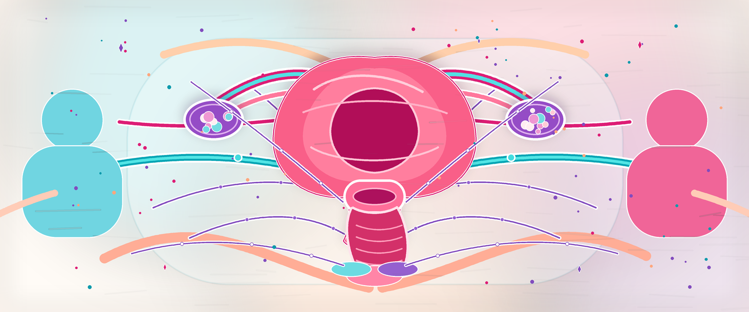
Female Sexually Important Organs
Start with the simplest frame: there are structures you can see (the external vulva), structures that lie beneath them (erectile bodies, glands, muscles, nerves and vessels), and the function those layered structures together produce (sensation, vasocongestion, lubrication, accommodation and orgasm). This chapter ascends in exactly that order — surface map first, then the tissues underneath, then the nerves, blood and lymph that serve them, then the integrated sexual response, and finally the consultation. Each section assumes only what came before it.
Once the parts are in place, the key insight is that female sexual anatomy is not a list of visible parts. It is an integrated system of erectile tissue, somatic sensation, autonomic vasocongestion, vestibular mucosa, oestrogen-responsive epithelium, pelvic-floor relaxation, lubrication, blood flow, lymph drainage and psychological safety. O&G candidates need this anatomy for dyspareunia, vulvodynia, postpartum pain, menopause, Bartholin disease, vulval dermatoses, sexual trauma, gender-based violence, FGM/C, vulval surgery and counselling after childbirth.
The first rule is precision without judgement. Normal vulval appearance varies widely. The aim of anatomy is to localise pain, explain function, avoid injury and examine respectfully, not to define a cosmetic norm.
External Map: Vulva, Vestibule and Perineum
The vulva includes the mons pubis, labia majora, labia minora, clitoral complex, vestibule, urethral meatus, vaginal introitus, posterior fourchette and perineal body. The vestibule is the mucosal space between the labia minora containing the urethral and vaginal openings. This distinction matters because vestibular pain, vulval skin disease and vaginal pain are not the same anatomical problem.
| Structure | Tissue type | O&G relevance |
|---|---|---|
| Mons pubis | Hair-bearing fat pad over pubic symphysis | Pubertal change, folliculitis, scars |
| Labia majora | Hair-bearing folds with fat, glands and vascular tissue | Swelling, dermatoses, trauma, vulval masses |
| Labia minora | Hairless folds, variable size and pigmentation | Normal variation, fissures, pain, FGM/C anatomy |
| Vestibule | Non-keratinised mucosa between labia minora | Vestibulodynia, discharge, urethral/Bartholin openings |
| Posterior fourchette | Posterior labial joining point | Fissuring, childbirth trauma, dyspareunia |
| Perineal body | Fibromuscular node between vagina and anus | Sexual pain after scar, posterior support, obstetric tears |
Several exam mistakes come from using vague words. "Vaginal pain" might mean vestibular burning at the introitus, posterior fourchette fissuring, levator spasm, deep uterosacral pain, bladder pain or true vaginal mucosal disease. A good answer localises the tissue and the nerve territory.
Embryology, Homology and Normal Variation
The external genitalia have shared embryological origins before sexual differentiation. This matters in DSD assessment, paediatric counselling and respectful adult examination because many structures are homologous rather than "extra" or "abnormal".
| Embryological structure | Female derivative | Clinical use |
|---|---|---|
| Genital tubercle | Clitoris | Explains clitoral enlargement and androgen-effect questions |
| Urogenital folds | Labia minora | Helps describe labial fusion, FGM/C effects and normal asymmetry |
| Labioscrotal swellings | Labia majora | Explains hair-bearing outer folds and inguinal-labial swelling pathways |
| Urogenital sinus/vestibular region | Vestibule, lower-vaginal contribution, urethral area | Separates vestibular symptoms from vaginal-canal symptoms |
Normal adult vulvas vary in labial length, pigmentation, rugosity, clitoral hood coverage and vestibular colour. A Primary answer should therefore avoid cosmetic language. Use terms such as enlarged, oedematous, fused, scarred, ulcerated, fissured, tender, atrophic, erythematous or mass-like only when those are anatomical findings.
Clitoral and Vestibular Erectile Anatomy
The clitoris is an internal-external erectile organ. The visible glans is only the distal part. The full clitoral complex includes the glans, body, crura and neurovascular structures; the vestibular bulbs are paired erectile tissues closely related to the introitus and clitoral bodies.
| Component | Anatomy | Clinical relevance |
|---|---|---|
| Glans clitoris | Visible distal structure, densely innervated, covered partly by prepuce | High sensory density; vulnerable in vulval surgery, trauma and FGM/C |
| Body | Paired corpora cavernosa joined anteriorly | Deep erectile tissue; not visible on inspection |
| Crura | Erectile roots attached to ischiopubic rami | Relationship to ischiocavernosus and pubic arch |
| Prepuce/hood | Fold over glans | Adhesions, dermatoses, scarring, FGM/C effects |
| Vestibular bulbs | Paired erectile tissue flanking vaginal opening, deep to labia minora | Engorgement, haematoma, episiotomy/tear proximity |
During arousal, increased blood flow fills erectile tissue and venous outflow is partly restricted by surrounding tissues and muscles. The bulbospongiosus and ischiocavernosus muscles help compress erectile tissues. This produces genital swelling, increased sensitivity and lubrication through vascular transudation.
Mechanism-to-clinical-consequence chain:
| Mechanism | Tissue effect | Possible symptom |
|---|---|---|
| Pudendal neuropathy or dorsal clitoral nerve injury | Reduced somatic sensation | Reduced clitoral sensation or neuropathic pain |
| Vestibular bulb trauma | Venous bleeding in loose tissue | Painful vulval haematoma |
| Scarring over clitoral prepuce or glans | Tethering, exposure or concealment | Pain, altered sensation, hygiene difficulty |
| Pelvic-floor overactivity around introitus | Compression and guarding | Entry dyspareunia, examination intolerance |
Avoid the reductionist statement that sexual function is "clitoral only" or "psychological only". Sexual response requires intact tissue, blood flow, nerves, hormones, pelvic-floor relaxation, context, consent and absence of pain.
Sexual Response as Anatomy in Motion
Sexual response is a neurovascular event layered onto mucosa and pelvic floor. Parasympathetic activity supports vasocongestion and lubrication. Somatic pudendal pathways provide touch, pain and motor control of perineal muscles. Sympathetic and visceral pathways contribute to orgasm, uterine/cervical sensation and some deep pelvic pain.