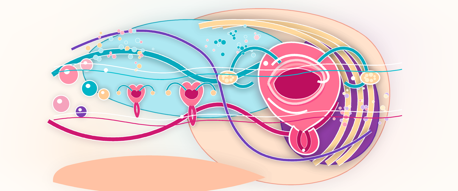
Female Child Genital Tract
Clinical Overview
The child's genital tract is not a miniature adult tract. It is a developmental stage with its own surface anatomy, epithelial biology, proportions, microbial environment, examination technique and safeguarding context. To read it correctly you need three layers, learned in order: first how the tract was built in the embryo, because every structural anomaly is a fault somewhere along that build; second how the same finished structures behave under different hormone states, because the dominant variable in childhood is not size but oestrogen state; and third how those two facts explain the clinical presentations you will actually meet.
The hormone story sets the whole frame. The newborn may briefly show maternal-oestrogen effects that wash out over the first weeks. The prepubertal child is then hypo-oestrogenic for years. The pubertal adolescent becomes progressively oestrogenised and her anatomy converges on the adult reproductive pattern. The same symptom therefore means different things at different ages: a little neonatal mucus or a small withdrawal bleed can be physiological; prepubertal discharge usually reflects an exposed, thin, neutral-pH vulvovaginal surface rather than adult candidiasis; cyclic pain without menses points to outflow anatomy; and a virilised newborn demands structured difference-of-sex-development (DSD) thinking and urgent endocrine assessment. Genital trauma or a sexually transmitted infection in a child must be interpreted through safeguarding law and expert examination pathways, but the anatomical description itself must stay precise and unemotional.
High-yield chains, which the rest of this chapter unpacks:
Chromosomal sex -> gonadal development -> hormone production and action -> internal ducts and external genitalia; a DSD is a mismatch somewhere along this sequence.
Low oestrogen -> thin squamous epithelium, little glycogen, neutral pH -> less lactobacillus protection -> irritation and non-specific vulvovaginitis.
Small labia majora, no pubic hair, short ano-vulval distance -> poor mechanical protection -> faecal contamination and irritant exposure.
Oestrogen at puberty -> thick glycogenated vaginal epithelium -> lactobacillus metabolism -> acidic adult-type vaginal environment.
Outflow obstruction at the hymen, distal vagina or a transverse septum -> menstrual blood cannot drain -> haematocolpos -> cyclic pain, mass effect, urinary retention or constipation.
Core Knowledge
How the Tract Is Built: Embryology First
Start with the build, because the obstructions and the DSDs you will meet are all faults in this sequence. Around the fifth to sixth week the embryo reaches an indifferent stage: it carries both duct systems and an undifferentiated gonad, and the external genitalia of a male and a female are indistinguishable. The whole female tract is then produced by what does, and does not, happen next.
Gonad. The gonad has a triple origin: coelomic epithelium of the genital ridge, the underlying mesenchyme, and the primordial germ cells that migrate in from the yolk sac wall via the hindgut and dorsal mesentery, reaching the ridge at about four weeks. The switch is the SRY gene on the short arm of the Y chromosome, which drives the transcription factor SOX9. With SRY present, the supporting cells become Sertoli cells and a testis forms from about seven weeks. Without SRY the default pathway is ovarian: the same four lineages become granulosa cells, oogonia, thecal cells and ovarian stroma, and the ovary becomes identifiable at about ten weeks.
Ducts. Two paired ducts run side by side: the paramesonephric (Müllerian) ducts and the mesonephric (Wolffian) ducts. Their fate is decided by two testicular products, so the absence of those products is what builds a female tract:
- The fetal testis secretes anti-Müllerian hormone (AMH) from Sertoli cells, which regresses the Müllerian ducts, and testosterone from Leydig cells, which stabilises the Wolffian ducts.
- In a female there is no AMH, so the Müllerian ducts persist and form the fallopian tubes, uterus, cervix and upper vagina. There is no testosterone, so the Wolffian ducts regress.
The Müllerian ducts grow caudally, reach the dorsal wall of the urogenital sinus by about nine weeks, and their lower portions fuse in the midline. Mesoderm proliferating around the fused segment forms the muscular walls of the uterus and cervix during the fourth month. The vagina is a fusion zone: where the Müllerian ducts meet the urogenital sinus they induce a solid vaginal plate, made partly of sinus endoderm and partly of Müllerian tissue, which then canalises at about sixteen to eighteen weeks to open the vaginal lumen. The upper vagina is therefore Müllerian and the lower vagina and vestibule derive from the urogenital sinus — a single embryological fact that explains why obstruction can sit at the hymen, in the distal vagina, or higher up.
External genitalia. The genital tubercle, the urogenital (genital) folds and the labioscrotal swellings are present by about six weeks but look identical in both sexes until roughly ten weeks. In the female, in the absence of androgen action, the genital tubercle stays small and becomes the clitoris, the genital folds become the labia minora, and the labioscrotal swellings become the labia majora. In the male the same primordia, driven by testosterone converted to dihydrotestosterone (DHT) by 5α-reductase, become penis and scrotum. This shared origin is why excess androgen in a 46,XX fetus virilises the external genitalia, and why impaired androgen synthesis or action in a 46,XY fetus leaves them female-appearing.
| Female structure | Embryological origin | Driver / absence of driver |
|---|---|---|
| Ovary | Genital ridge, no SRY | Default pathway when SRY absent |
| Tubes, uterus, cervix, upper vagina | Paramesonephric (Müllerian) ducts | Persist because no AMH |
| Lower vagina, vestibule | Urogenital sinus | Endodermal contribution to the vaginal plate |
| Clitoris | Genital tubercle | Stays small without androgen/DHT |
| Labia minora | Genital (urogenital) folds | Do not fuse without androgen |
| Labia majora | Labioscrotal swellings | Do not fuse to a scrotum without androgen |
Hold one more fact from this section: the urinary and genital systems are built together from the same urogenital ridge, so a Müllerian anomaly should always prompt a question about the kidney.
The Ovary and Its Germ Cell Stock
The child's ovary already carries her entire reproductive future, decided before birth. From early gestation the oogonia enter meiosis and arrest at the first meiotic prophase (diplotene); each arrested oocyte surrounded by a single flat layer of pregranulosa cells is a primordial follicle. The germ-cell stock peaks at roughly 6–7 million around 20 weeks, then falls through atresia to about 1–2 million at birth, and to roughly 300,000–600,000 by puberty. No new oocytes are made after birth — this finite, declining pool is the basis of the entire ovarian reserve concept the trainee will meet again in fertility work.