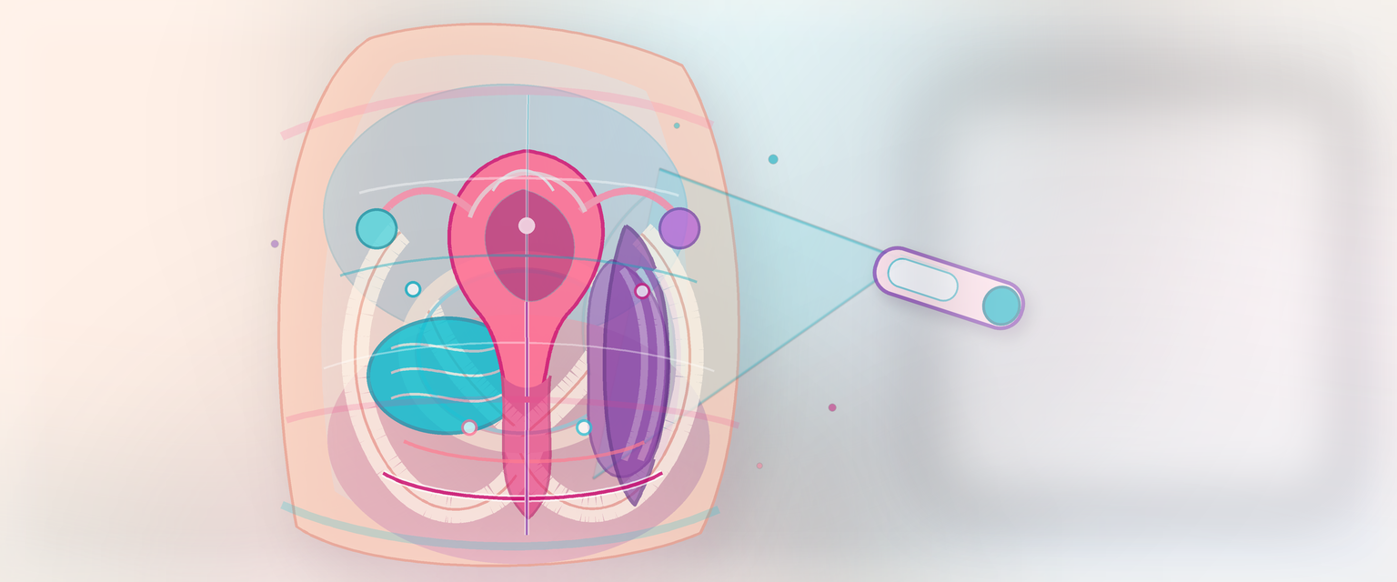
Surface Anatomy of the Female Abdomen and Pelvis
Surface anatomy is the translation layer between the woman in front of you and the structures hidden under skin, fat, muscle, peritoneum and pelvic fascia. In O&G it tells you where pain is being felt, where the uterine fundus should be, where to place a laparoscopic port, why a caesarean scar is risky on re-entry, how to examine the groin, where a pudendal block is aimed, and how to communicate an abdominal or pelvic finding without vague language.
The point is not to memorise a drawing. The point is to build a three-dimensional map: skin landmark -> abdominal wall layer -> peritoneal cavity or extraperitoneal plane -> pelvic organ, vessel, nerve or lymph route.
Before any of that, fix one mechanical idea that every later section rests on. The trunk is a muscular cylinder slung between two bony rings — the costal margin above and the pelvic ring below — splinted apart by the vertebral column. The top ring is sealed by the diaphragm; the bottom ring is sealed by the pelvic floor. Abdominal wall muscle keeps a gentle positive intra-abdominal pressure (around +5 mmHg at rest) and the diaphragm closes the top, so a problem in one compartment is felt and transmitted into the next. That single picture explains why diaphragmatic blood gives shoulder-tip pain, why a raised pregnant diaphragm changes where chest and abdominal injuries land, and why pressure and contents shift the moment you insufflate at laparoscopy. Everything that follows is just that cylinder, examined layer by layer.
Figure A2.1 — The nine abdominal regions are coordinates, not diagnoses: name where pain is felt (with correct left/right), then reason about what lies beneath — right iliac fossa ectopic/torsion/PID, epigastric/RUQ HELLP, shoulder-tip from diaphragmatic irritation.
Fixed Landmarks Before Variable Findings
Start every surface-anatomy answer with fixed bony landmarks. Soft tissues move with pregnancy, obesity, bladder filling, bowel distension, fibroids, ascites and surgical scars; bones are the framework.
| Surface landmark | Deep relation | O&G use |
|---|---|---|
| Xiphisternum | Lower sternum, diaphragm nearby | Late fundal-height orientation; upper abdominal pain context |
| Costal margin | Upper abdomen, liver and spleen deep to it | RUQ pain in pre-eclampsia/HELLP; trauma in pregnancy |
| Umbilicus | Variable; often around L3/L4 in adults | Port planning, abdominal quadrants, fundal-height reference |
| Iliac crests | Intercristal plane near L4 | Neuraxial-landmark context; abdominal wall orientation |
| Anterior superior iliac spine | Lateral inguinal ligament attachment | Groin anatomy, port triangulation, ilioinguinal region |
| Pubic symphysis | Anterior pelvic ring, retropubic space | Caesarean entry, bladder reflection, suprapubic pain |
| Pubic tubercle | Medial inguinal ligament attachment | Inguinal canal, femoral canal, groin-node examination |
| Ischial spine | Midpelvis, sacrospinous ligament | Fetal station, pudendal block, sacrospinous fixation danger zone |
| Ischial tuberosity | Inferior pelvis | Perineal triangle, episiotomy and pudendal canal orientation |
Pregnancy changes this map. The enlarging uterus rises out of the pelvis after the first trimester, displaces bowel upward and laterally, stretches the abdominal wall, changes the umbilicus, raises the bladder reflection in the lower abdomen and makes a standard port or incision less standard. Previous surgery changes it again: a skin scar does not tell you where the fascia was opened, whether the bladder is adherent, or where bowel is stuck to the anterior wall.
Planes That Anchor Safe Examination
Surface planes are useful only if they help you localise risk. In O&G, they mainly orient upper abdominal pain, neuraxial estimates, port placement and the changing position of non-gynaecological organs in pregnancy.
| Plane or point | Approximate level | Use |
|---|---|---|
| Transpyloric plane | Around L1, a handbreadth below xiphisternum | Upper abdominal orientation: pylorus, renal hila and pancreatic neck region |
| Subcostal plane | Around L3 | Separates upper from central abdomen; helps RUQ and flank localisation |
| Intercristal plane | Around L4 | Aorta has divided; useful but imperfect neuraxial and vascular orientation |
| McBurney-type point | Line from umbilicus to right ASIS | Non-pregnant appendix clue; pregnancy displaces caecum and appendix upward |
| Langer/tension lines | Transverse in lower anterior abdominal wall | Explains why transverse incisions often heal with less visible scarring |
The trap is treating these as fixed coordinates in every patient. Obesity, prior surgery, a gravid uterus, fibroids, ascites and a distended bladder all change the surface-to-deep relationship. In late pregnancy, appendicitis may localise above the right iliac fossa and an incision should follow the clinical maximum tenderness rather than a memorised non-pregnant point.
Dermatomes and Referred Pain: the Hidden Layer of the Map
Surface anatomy is not only where structures sit; it is where their pain is felt. The anterior abdominal wall is supplied segmentally by the lower thoracic and upper lumbar nerves, so a visceral structure refers pain to the dermatome of its own segmental nerve supply rather than to the spot it actually occupies. Learn this map before you interpret any pain stem, because it is what lets you reason about a pelvic organ from a chest- or flank-level symptom.
| Dermatome / referral | Surface area | O&G meaning |
|---|---|---|
| T7–T9 | Epigastrium | Upper-gut and gallbladder referral; HELLP liver-capsule pain sits here |
| T10 | Umbilical band | Ovary and tube refer to T10–T11; early periumbilical visceral pain (e.g. appendix) before it localises |
| T11–L1 | Lower abdomen, groin, mons | Body of uterus and round ligament; lower-segment and round-ligament pain |
| S2–S3 | Deep pelvic / perineal | Cervix and upper vagina; cervical and deep pelvic visceral pain |
| Obturator (L2–L4, mainly L3) | Medial thigh | Pelvic peritoneum and ovary irritating the obturator nerve refer pain down the inner thigh |
| Phrenic (C3–C5) | Shoulder tip | Diaphragmatic irritation by intraperitoneal blood, pus or gas |
Two clinical consequences follow directly. First, ovarian or pelvic-peritoneal pathology can present as medial-thigh pain because the obturator nerve runs on the lateral pelvic wall and its L3 fibres also supply the pelvic peritoneum — a useful sign in a torsion or a tubo-ovarian abscess. Second, chest pathology can masquerade as an abdominal event: the parietal pleura is supplied by the intercostal nerves, so lower-lobe pleural irritation refers pain to the same dermatome on the anterior abdominal wall. In pregnancy the diaphragm is pushed up, which both raises the spread of referred pain and means a chest injury is more likely to involve intra-abdominal organs — and it shifts the safe site for a chest drain up one intercostal space.

Figure A2.2 — Dermatomes and referred pain: pain is felt on the surface map of the supplying spinal segment, not where the organ sits — T10 umbilical, T11–L1 lower abdomen/mons, S2–S3 perineum, obturator to the medial thigh, and diaphragmatic blood to the shoulder tip.
Abdominal Regions: Coordinates, Not Diagnoses
The abdomen is commonly divided into four quadrants or nine regions. Either system is useful if you remember that a region is a coordinate, not a diagnosis.
| Region or pain site | O&G possibilities | Important mimics |
|---|---|---|
| Right iliac fossa | Ectopic pregnancy, ovarian torsion, ruptured cyst, PID, endometriosis | Appendicitis, ileitis, ureteric colic |
| Left iliac fossa | Ovarian cyst, torsion, PID, endometriosis | Diverticulitis, constipation, ureteric colic |
| Suprapubic | Early pregnancy loss, labour, uterine tenderness, bladder pain | Cystitis, retention, pelvic fracture |
| Epigastric/RUQ | Severe pre-eclampsia, HELLP liver capsule pain | Gallstones, hepatitis, reflux, pancreatitis |
| Flank/loin | Pregnancy hydronephrosis, pyelonephritis, ureteric obstruction | Renal colic, musculoskeletal pain |
| Generalised abdomen | Haemoperitoneum, sepsis, rupture, peritonitis | Bowel obstruction, perforation, pancreatitis |
| Shoulder-tip pain | Diaphragmatic irritation from intraperitoneal blood or gas | Thoracic pathology, gallbladder disease |
In acute pelvic pain, put the symptom onto three maps at once:
- Surface map: where is the pain felt?
- Organ map: which pelvic structures lie deep or refer there?
- Peritoneal and autonomic map: is this visceral stretch, somatic peritoneal irritation, diaphragmatic irritation, or myofascial pain?
A ruptured ectopic pregnancy may give pelvic pain, shoulder-tip pain and collapse because intraperitoneal blood irritates the peritoneum and diaphragm while blood loss drives shock. Ovarian torsion may give iliac fossa pain and vomiting because visceral afferents and autonomic response are involved. Chronic pelvic pain is even less one-organ in nature; modern reviews emphasise musculoskeletal, visceral, neuropathic and centrally mediated contributors, so a surface pain point should start an examination, not end it.