In one line
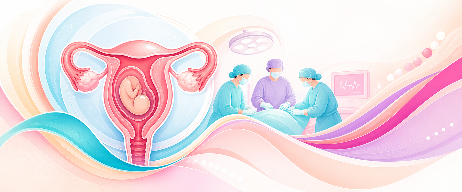
Uterine rupture is a full-thickness breach of the uterine wall threatening exsanguination of the mother and acute hypoxia of the fetus; survival is governed almost entirely by decision-to-delivery time, so the management plan is recognition → resuscitate → deliver immediately (laparotomy) → arrest haemorrhage → make a definitive repair-versus-hysterectomy decision in theatre.
This chapter assumes the mechanism, classification (complete vs dehiscence/window) and risk-factor groundwork from the Intermediate course — see uterine rupture basics and instrumental delivery — and concentrates on the consultant-level plan, the appraisal of who may labour, and the unresolved controversies.
Subtypes — why the distinction changes the plan
The word "rupture" hides four clinically distinct lesions, and the management diverges at the first branch point. The basic complete-vs-dehiscence split is covered in Intermediate; the consultant-level question is which one you are dealing with and what each one demands.
- Scar dehiscence (the "window"). A bloodless separation of the myometrium of a prior scar with the visceral peritoneum/serosa intact, the fetus and membranes still contained, and no significant haemorrhage. This is usually an incidental finding at repeat caesarean or a quiet ultrasound thinning — it is not an emergency, does not cause the rupture triad, and in an asymptomatic woman at elective caesarean needs only inspection and a clean two-layer closure. Conflating a window with a rupture is the commonest classification error and leads to over-treatment; the reverse error — dismissing pain in a labouring scarred woman as "just a window" — is the lethal one.
- Complete rupture of a scarred uterus (lower-segment). Full-thickness disruption usually along the prior transverse lower-segment scar, with communication into the peritoneal cavity. Because the lower segment is relatively avascular and the tear follows a predictable transverse line, haemorrhage is often more controllable and the tissue planes are cleaner — this is the subtype most amenable to primary repair. It is also the subtype where the herald is a subtle CTG change rather than collapse, because the scar gives way before the great vessels are reached.
- Complete rupture of an unscarred uterus. A different disease. The tear is unpredictable in site and shape — typically a vertical or stellate fundal/lower-segment tear driven by obstructed labour or uterotonic over-stimulation — frequently extends laterally into the broad ligament and uterine vessels, and presents later and more catastrophically (collapse, fetal extrusion, massive haemoperitoneum). Devitalised, ragged edges and vascular extension make this the subtype that most often forces hysterectomy. In SA district practice this is the high-parity, under-monitored, obstructed-labour patient — the rupture you must actively look for because there is no scar to "blame".
- Atypical sites — classical/vertical scar, posterior, and lateral/cervical extensions. A prior classical or inverted-T/J upper-segment scar ruptures higher, earlier, and often before labour through the contractile fundus, where bleeding is brisk and repair is harder; this is why a classical scar prohibits labour outright. Lateral and cervical/vaginal extensions threaten the uterine artery, ureter and bladder and convert a "simple" repair into vascular surgery. Identifying the subtype is what lets you anticipate the operative difficulty before you open.
The mechanism→consequence links are: a scarred lower-segment tear → cleaner plane, controllable bleed, repair feasible; an unscarred or lateral tear → vascular/ureteric jeopardy, devitalised tissue, hysterectomy more likely; a classical/posterior tear → early, pre-labour, brisk — surveillance and prohibition of labour, not detection in labour, is the only defence.
Assessment
Rupture is a clinical diagnosis confirmed at laparotomy; do not wait for imaging. The classic triad (pain, bleeding, non-reassuring fetal status) is unreliable, so weight the early, sensitive signs.
- Fetal heart rate is the most consistent herald. An abnormal CTG — typically prolonged or recurrent deceleration / bradycardia — precedes or accompanies most ruptures and is present in the majority of cases. New, atypical variable or late decelerations in a woman labouring with a scar should be read as rupture until disproved.
- Maternal signs: scar/abdominal pain breaking through effective regional analgesia, cessation of previously efficient contractions, vaginal bleeding, haematuria, loss of station / receding presenting part, palpable fetal parts abdominally, and signs of hypovolaemia (tachycardia, hypotension, restlessness). Shoulder-tip pain signals haemoperitoneum.
- In an unscarred uterus (multiparity, obstructed labour, injudicious oxytocin, prior perforation/myomectomy) rupture is often more catastrophic and later-recognised — a key SA reality in high-parity, under-monitored district labour.
- Investigations are resuscitative, not diagnostic: group-and-crossmatch (≥4 units), FBC, coagulation/fibrinogen, U&E — but never let a request for a scan or bloods delay laparotomy when the picture fits.
The advanced read — atypical presentations and the judgement calls
The basic triad is covered at Intermediate; the harder cases are the ones that do not present classically.
- The silent epidural dehiscence. Under a well-functioning epidural, a labouring scarred uterus can rupture with no maternal pain — the only sign is the CTG and a rising baseline maternal pulse. A "top-up that doesn't work" or breakthrough scar pain through a previously perfect block is a red flag, not an analgesia failure. The lesson: in a scarred labour, the CTG plus the maternal heart-rate trend outrank the pain history.
- The postpartum rupture. Rupture can declare after a successful VBAC delivery — persistent lower-abdominal pain, ongoing fresh bleeding with a contracted fundus, unexplained tachycardia, or a fall in haemoglobin not explained by the visible loss. Do not anchor on uterine atony; a contracted uterus that keeps bleeding, or a woman who is "shocked out of proportion" to the measured loss, needs the abdomen examined and imaged or explored.
- Loss of station as a hard sign. A presenting part that recedes on vaginal examination (the fetus retracting into the abdomen) is close to pathognomonic and trumps a "reassuring" cervix.
- Severity stratification at the bedside. The variables that predict death and hysterectomy — and therefore the urgency and the call for blood and a second surgeon — are complete (vs dehiscence) extent, fetal extrusion/placental separation, lateral/broad-ligament extension, unscarred uterus, and time elapsed since the FHR change. A woman with a receding presentation, abdominal fetal parts and a fetal bradycardia is at the catastrophic end and should be in theatre while you are still drawing blood; a quiet window found at elective section is at the benign end.
- Where investigations mislead. A bedside ultrasound may show free fluid or an empty uterus with the fetus in the abdomen, but a normal scan never excludes rupture and the time spent obtaining it is the time the baby does not have. CTG is sensitive but not specific — the same prolonged deceleration occurs in abruption and cord prolapse, which is why the response (immediate delivery) is identical and you do not need to discriminate them before acting. Haematuria points to bladder/anterior-segment involvement but its absence proves nothing.
Investigations
There is no diagnostic test to wait for. The investigations earn their place by driving resuscitation and the surgical plan, not by confirming the diagnosis — that is made with a knife.
- Group-and-crossmatch ≥4 units, FBC, fibrinogen/coagulation, U&E, venous lactate/base deficit. Lactate and base deficit index the degree of shock and the resuscitation debt better than a single blood pressure; a rising lactate in a "stable" woman is occult haemorrhage. Fibrinogen is the first clotting factor to fall in obstetric haemorrhage — a level <2 g/L in this setting is ominous and pre-empts cryoprecipitate/fibrinogen concentrate (postpartum-haemorrhage).
- Point-of-care haemoglobin and, where available, viscoelastic testing (ROTEM/TEG) to direct goal-directed product replacement rather than fixed ratios — useful at tertiary level, absent at most district hospitals, where you replace empirically and clinically.
- Imaging has an antenatal, predictive role — not an intrapartum, diagnostic one. This is the one place a number helps: sonographic lower-uterine-segment (LUS) thickness has been studied to triage who may safely labour after caesarean. The Kok meta-analysis (2013) supports antenatal LUS measurement for predicting a uterine defect, but cutoffs are method-dependent and unstandardised — reported thresholds span roughly 1.5–4.05 mm, with a full LUS thickness below ~2.3 mm and a myometrial thickness around ~2 mm marking higher risk (a myometrial cutoff of ≤2 mm has been reported with sensitivity ~91.7% and specificity ~81.8% for a uterine defect in labour). The honest interpretation is that LUS thickness is a research/counselling adjunct that is not validated for routine clinical decisions, the measurement is operator- and route-dependent (transvaginal reads thinner than transabdominal), and it must never override the clinical decision to deliver. It does not exist in most SA district workflows.
- VBAC success-prediction models belong in the antenatal clinic, not the labour ward: the Grobman/MFMU VBAC calculator estimates the probability of vaginal birth from maternal factors and informs counselling and selection (the 2021 revision removed race/ethnicity after the original 2007 model was shown to systematically under-predict success in Black and Hispanic women — a relevant equity point for SA). It predicts success, not rupture, and a high predicted-success score does not licence aggressive augmentation.
Differential diagnosis
The differentials matter only insofar as they change the immediate action — and for the catastrophic intrapartum picture, most of them do not, because the response (resuscitate + deliver) is shared.
- Placental abruption — pain, bleeding, non-reassuring fetal status and a tense uterus; in a scarred labouring woman the two are clinically indistinguishable and the management (immediate delivery + haemorrhage control) is identical, so do not delay theatre to tell them apart. They also coexist: rupture can cause placental separation.
- Cord prolapse / vasa praevia — sudden fetal bradycardia; excluded on examination, and again the answer is immediate delivery.
- Severe uterine atony / placenta accreta spectrum as a cause of postpartum collapse — distinguished by a boggy (atony) versus contracted-but-still-bleeding (rupture/extension) fundus; the contracted-uterus-with-ongoing-bleeding pattern is the one that must trigger examination of the lower segment and consideration of rupture rather than escalating uterotonics.
- Broad-ligament haematoma without free rupture — pain and shock with concealed loss; found at laparotomy, managed by the same haemostatic principles.
- The intra-abdominal mimics (ruptured rudimentary horn, ruptured ectopic in a coexisting early pregnancy, splenic/hepatic pathology) — rare, but the unifying principle holds: an unstable obstetric abdomen with fetal compromise is explored, not investigated to exhaustion first.
The single management-changing distinction is dehiscence/window (observe, no emergency) versus complete rupture (theatre now) — the rest converge on the same resuscitate-and-deliver pathway.
Management
Management runs immediate → ongoing → long-term.
Immediate (the first minutes — run in parallel, not in sequence):
| Action | Detail (SA/SAMF) |
|---|---|
| Call for help | Declare obstetric emergency: senior obstetrician, anaesthetist, theatre, neonatology, blood bank, porter. In a district setting, activate transfer and prepare to operate locally — do not wait for a referral bed. |
| Resuscitate | Two large-bore cannulae, warmed crystalloid (Ringer's lactate), activate massive transfusion protocol, crossmatched then O-negative blood, tranexamic acid 1 g IV. |
| Stop the stimulus | Discontinue oxytocin immediately. |
| Deliver | Immediate laparotomy. This is a Category-1 (crash) caesarean; the fetus is delivered through the rupture or a fresh incision, then the bleeding is controlled. |
Ongoing (at laparotomy):
- Deliver the fetus and placenta, exteriorise the uterus, and identify the full extent of the defect. Control bleeding with clamps/pressure while the anaesthetist catches up on volume.
- Repair vs hysterectomy is the central judgement. Favour two-layer primary repair when the tear is a clean lower-segment scar dehiscence, the edges are viable, haemostasis is achievable, and future fertility matters. Favour hysterectomy when there is an extensive/stellate tear, broad-ligament or cervical/vaginal extension, uncontrollable haemorrhage, or devitalised tissue — do not persist with repair in an unstable, coagulopathic woman.
- Map adjacent structures: assess bladder (lower-segment/anterior tears), and exclude broad-ligament haematoma and ureteric injury before closing. Lateral extensions risk the uterine vessels and ureter.
- Manage the haemorrhage on its own track — this is postpartum-haemorrhage physiology: balanced resuscitation, fibrinogen-guided product replacement, and a low threshold for interventional radiology/internal iliac ligation where available.
Subtype-specific surgery and the operative judgement
The "repair vs hysterectomy" decision is the central operative judgement. Decide on four axes, in order: haemodynamic/coagulation state, extent and viability of the tear, vascular/ureteric involvement, and the woman's fertility wishes — and let the patient's stability veto everything else.
- Clean lower-segment scar rupture, stable woman, fertility desired → repair. Debride non-viable edges minimally, achieve haemostasis, and close in two layers (the single-layer/two-layer debate from Bujold applies to the index caesarean's recurrence risk, but at repair a secure double layer is standard). Inspect the bladder and run the uterine angles to exclude lateral extension before you are satisfied.
- Lateral / broad-ligament / cervical extension → control the vessels first. Open the broad ligament, identify and protect the ureter, secure the uterine artery, and only then decide if a repair is reconstructable. This is where uterine-artery or internal iliac (anterior division) ligation buys time and where over-confident suturing causes ureteric injury. A stellate or low cervical extension into the vagina that you cannot reconstruct cleanly is a hysterectomy.
- Unscarred, stellate, devitalised, or coagulopathic → hysterectomy, and do not delay it. Persisting with repair in an unstable woman is "a hysterectomy delayed and a death." Subtotal hysterectomy is faster and safer when the bleeding is from the body of the uterus; a total hysterectomy is needed when the tear extends into the cervix or the bleeding source is the lower segment/angles. Speed and the bleeding source dictate subtotal vs total.
- Damage control for the in-extremis patient. If the woman is cold, acidotic and coagulopathic (the "lethal triad"), the correct operation is damage control: aortic compression or cross-clamping, pelvic packing, rapid haemostatic control, leave-and-return — resuscitate in ICU and return to definitive surgery once warm and clotting. Heroically completing a perfect anatomical repair on a dying woman is the wrong instinct.
- Adjuncts and the SA reality. Tranexamic acid 1 g IV early (the WOMAN-trial principle — give it within the first hours) is cheap, EML-listed and applies to the haemorrhage; balloon tamponade does not control a rupture (it controls atony) and is the wrong tool here; interventional radiology and cell salvage exist at tertiary centres only. At a district hospital the realistic kit is clamps, ligation, packing, a second pair of hands, and an early transfer-or-call decision — which is why the systems answer (operate locally, do not wait for a referral bed) is the safe one.
Long-term:
- Counsel explicitly on the next pregnancy. After a prior rupture, elective repeat caesarean before labour is mandated (recurrence risk is materially higher than a first VBAC attempt), typically at 36–37 weeks to pre-empt labour; planned VBAC is contraindicated. A previous classical (upper-segment) scar carries the same prohibition.
- Debrief the woman and team; complete a structured adverse-event review and, in SA, an MMR/PPIP perinatal-loss audit if the baby died.
Postnatal, recurrence and counselling in depth
The episode is not closed when the bleeding stops. The consultant owns the recovery, the future-fertility conversation, and the audit.
- Immediate postnatal. Critical-care-level monitoring for ongoing concealed bleeding (serial haemoglobin, urine output, abdominal girth/pain), thromboprophylaxis once haemostasis is secure (these women are both bleeding- and clotting-risk — balance it deliberately), and proactive treatment of the consequences of massive transfusion (hypocalcaemia, hyperkalaemia, dilutional coagulopathy). Where the baby died or was severely asphyxiated, structured bereavement support and honest, documented disclosure.
- Recurrence counselling — give a range, not false precision. Published recurrence of rupture in a subsequent pregnancy varies widely across small series (figures from a few percent up to the high teens, and some recent cohorts report none) — the honest message is that the risk is real and materially higher than a primary scar, that it tends to recur earlier in gestation (often before labour), and that the consequence (not just the probability) is what drives the plan. The plan is therefore the same regardless of the exact number: no labour next time.
- Plan the next pregnancy concretely. Elective caesarean before the onset of labour — commonly 36–37 weeks, individualised earlier if the prior rupture was a fundal/classical tear that ruptured pre-labour — at a unit with theatre, blood and neonatal support immediately available; counsel against home or far-from-facility birth; document the prior tear site and repair in the discharge summary because it dictates the next delivery's timing. Where childbearing is complete, a fully informed discussion of sterilisation at the index repair or interval is appropriate but never coerced in the emergency.
- The systems loop. Every rupture is a perinatal/maternal audit event. In SA, enter it into PPIP/the perinatal-morbidity meeting, and a maternal death into the NCCEMD/Saving Mothers confidential enquiry; the avoidable factors that recur — inappropriate augmentation of a scarred or grand-multiparous labour, delayed recognition, no continuous monitoring, no on-site theatre — are systems failures the audit is meant to fix.
The evidence & the controversy
The defensible plan rests on three appraised pillars. First, who labours. Pooled data put symptomatic rupture in planned VBAC at roughly 0.5% (Landon's MFMU cohort 0.7%; Guise's AHRQ review 0.47% for trial of labour vs 0.03% for elective repeat caesarean), against a ~74% chance of vaginal birth. The honest counselling number is therefore small absolute risk, large consequence — and the consequence is what drives the conversation: in Landon, term hypoxic-ischaemic encephalopathy was 0.46/1000 in trial of labour and zero with elective repeat caesarean. That asymmetry, not the rupture rate alone, is the basis of individualised consent. The decisive factor is who is selected: a prior vaginal birth (especially a prior successful VBAC) is the single best predictor of success and lowers rupture risk; two prior low-transverse caesareans may be offered VBAC only after senior counselling; a classical scar is an absolute contraindication. Selection tools (the Grobman/MFMU calculator) inform the success side of the conversation but do not quantify rupture and do not licence augmentation.
Second, what raises the risk — and what is therefore controllable. Induction and augmentation are the modifiable culprits. The classic quantification is Lydon-Rochelle (NEJM 2001): compared with elective repeat caesarean without labour, spontaneous labour carried a relative risk of rupture of 3.3, induction without prostaglandins 4.9, and prostaglandin induction 15.6 (95% CI 8.1–30.0), with an absolute rupture rate of 24.5 per 1000 in the prostaglandin group. That single study is why misoprostol is contraindicated for induction with a previous caesarean (term), why prostaglandins are avoided, and why oxytocin is used cautiously, if at all. Al-Zirqi's 40-year Norwegian series independently tied rising rupture to oxytocin augmentation and prostaglandin induction. The SA NDoH position for district practice is to not augment a previous-caesarean labour and to conduct it only where caesarean is available 24-hours with continuous CTG. A short interdelivery interval (≤24 months; Bujold adjusted OR 2.65) and single-layer closure (OR 4.33) independently raise risk, informing both selection and the index caesarean's surgical technique. The wider induction appraisal — agents, mechanical methods and scar-specific cautions — is developed in the Final's induction-of-labour chapter.
Third — the genuinely decisive controversy — is time, not technique. Survival is set by the decision-to-delivery interval. Al-Zirqi's 2018 outcome series quantified it: delivery within 20 minutes limited intrapartum/infant death to ~10%, whereas >30 minutes carried an adjusted OR of ~16.7 for death, and placental separation/fetal extrusion an OR of ~17.9. An 18-minute target is widely cited. Rupture management is fundamentally a systems problem — drills, in-house theatre access, and a resuscitate-while-you-deliver mindset save babies more than any choice of suture. The corollary for prediction: antenatal LUS-thickness measurement (Kok 2013) and success calculators refine selection, but no antenatal test reliably predicts the individual rupture — which is exactly why the safety net is a system that can deliver in minutes, not a screening test.
Landmark trials & key evidence
| Trial (year) | Question | Key finding | What it changed |
|---|---|---|---|
| Landon MFMU (2004) | Maternal/perinatal outcomes of trial of labour vs elective repeat CS | Symptomatic rupture 0.7% in TOLAC; term HIE 0.46/1000 (0 with ERCS), 2 neonatal deaths | Anchored modern VBAC counselling on absolute risk + the neonatal-consequence asymmetry |
| Guise AHRQ review (2010) | Pooled rupture risk by mode | Rupture 0.47% TOLAC vs 0.03% ERCS; overall 0.30% | The quotable "~1 in 200" figure underpinning guideline counselling |
| Lydon-Rochelle (2001) | Does mode of labour onset change rupture risk after prior CS? (population cohort) | vs ERCS without labour: spontaneous RR 3.3, non-prostaglandin induction RR 4.9, prostaglandin induction RR 15.6 (8.1–30.0); 24.5/1000 absolute with prostaglandins | Grounded the prohibition on prostaglandin/misoprostol induction of a scarred uterus |
| Bujold (2002) | Effect of interdelivery interval and closure technique | ≤24 months adjusted OR 2.65; single-layer closure OR 4.33; ≤12 months rupture 4.8% | Drove interval-based selection and the two-layer-closure preference |
| Kok meta-analysis (2013) | Does antenatal LUS thickness predict uterine defect/rupture in TOLAC? | Thinner LUS predicts defect; cutoffs unstandardised (~1.5–4.05 mm), method-dependent; supports use as an adjunct, not a rule | Defined the (limited, non-routine) role of sonographic LUS measurement in selection |
| Al-Zirqi BJOG (2016) | 40-year trends in rupture (Norway) | Rose from 1.2 to 6.1 per 10,000; driven by oxytocin augmentation + prostaglandin induction | Implicated labour management, not just scar prevalence, in rising rupture |
| Al-Zirqi AJOG (2018) | Determinants of infant outcome after complete rupture | >30 vs <20 min to delivery OR 16.7 for death; placental separation/extrusion OR 17.9 | Made decision-to-delivery interval the central modifiable outcome lever |
| FIGO VBAC GPR (2025) | Contemporary good-practice VBAC framework | Rupture ~0.3–0.7%; success 60–80%; interbirth ≥18 months; facility readiness + continuous monitoring | Current global selection/counselling/safety standard |
Worked viva — how to structure the answer
Examiners give a stem like "a 31-year-old para 2, one previous caesarean, labouring at term at a district hospital, sudden fetal bradycardia, scar pain breaking through her epidural, and the presenting part has risen." A high-scoring answer runs:
- Frame it — "This is uterine rupture until proven otherwise: a scarred labouring woman with a fetal bradycardia, breakthrough scar pain and a receding presentation. I will not wait for a scan — the diagnosis is made at laparotomy."
- Resuscitate and deliver in parallel — "Declare a Category-1 emergency; call senior obstetrician, anaesthetist, theatre and neonatology; two large-bore lines, crossmatch ≥4 units, activate massive transfusion, tranexamic acid 1 g, stop the oxytocin — and move to immediate laparotomy. At a district hospital I operate locally rather than wait for a referral bed."
- State the operative plan and its branch points — "I deliver the fetus, identify the full extent of the tear, and decide repair versus hysterectomy on stability, extent, vascular/ureteric involvement and fertility wishes: two-layer repair for a clean lower-segment tear in a stable woman; hysterectomy for a stellate, devitalised, broad-ligament or coagulopathic picture; control the vessels and protect the ureter before any lateral repair; damage-control surgery if she is cold, acidotic and coagulopathic."
- Justify from evidence — "Decision-to-delivery time drives survival (Al-Zirqi 2018 — >30 min OR ~16.7 for death); the absolute rupture risk in planned VBAC is ~0.5% but the consequence is severe (Landon — HIE 0.46/1000, zero with elective caesarean); prostaglandin induction multiplies the risk (Lydon-Rochelle — RR 15.6), which is why misoprostol is contraindicated here."
- Anticipate complications — haemorrhage and coagulopathy, bladder/ureteric injury, broad-ligament haematoma, neonatal asphyxia, and a postpartum rupture presentation if she had delivered vaginally.
- Close the loop — "Next pregnancy: elective caesarean before labour at ~36–37 weeks, VBAC contraindicated; debrief the woman and family; enter it into PPIP and, if the baby died, the Saving Mothers/perinatal audit."
Exam traps & red flags
- Waiting for confirmation. Ordering a scan, a repeat CTG, or "watching for 10 minutes" in a scarred labouring woman with a new bradycardia is the classic fatal delay. The plan is laparotomy — the diagnosis is made in theatre.
- Forgetting the unscarred uterus. High-parity obstructed labour and over-stimulation rupture intact uteri; these are often the most catastrophic and the easiest to miss in a busy district unit.
- Mistaking a window for a rupture (and vice versa). An incidental dehiscence at elective caesarean needs inspection and closure, not an emergency; but dismissing breakthrough scar pain in labour as "just a window" is the lethal version of the same error.
- Reflex repair in the wrong patient. Persisting with repair in a coagulopathic, haemodynamically unstable woman with a stellate tear is a hysterectomy delayed — and a death. Know when to do damage control instead of a perfect repair.
- Missing the bladder, ureter or broad-ligament extension before closing. Always inspect adjacent structures and protect the ureter before any lateral repair.
- Trusting a normal scan or a "reassuring" LUS thickness. Imaging never excludes rupture, and LUS-thickness measurement is a counselling adjunct, not a clinical green light.
- Anchoring on atony postpartum. A contracted uterus that keeps bleeding, or shock out of proportion to visible loss, is a rupture/extension until the lower segment is examined — escalating uterotonics wastes time.
- Misadvising the next pregnancy. Offering VBAC after a prior rupture or classical scar is a hard error — these are absolute contraindications; plan elective caesarean before labour (≈36–37 weeks).
- Pharmacology trap: prescribing misoprostol/prostaglandin to induce a previous-caesarean labour. Contraindicated at term (Lydon-Rochelle RR 15.6). The same haemorrhage discipline applies as in caesarean-at-full-dilatation and major PPH.
Evidence anchors
- Landon MFMU, NEJM 2004 — DOI 10.1056/NEJMoa040405
- Guise AHRQ review, Obstet Gynecol 2010 — DOI 10.1097/AOG.0b013e3181df925f
- Lydon-Rochelle, NEJM 2001 — DOI 10.1056/NEJM200107053450101
- Bujold, AJOG 2002 — DOI 10.1067/mob.2002.127138
- Kok, Ultrasound Obstet Gynecol 2013 (LUS thickness meta-analysis) — DOI 10.1002/uog.12479
- Al-Zirqi, BJOG 2016 — DOI 10.1111/1471-0528.13394
- Al-Zirqi, AJOG 2018 — DOI 10.1016/j.ajog.2018.04.010
- FIGO good practice recommendations for VBAC, 2025 — DOI 10.1002/ijgo.70406
- RCOG Green-top Guideline No. 45, Birth After Previous Caesarean Birth (2015)
- SA NDoH — Guidelines for Maternity Care in South Africa / National Integrated Maternal and Perinatal Care Guideline (2024): previous-CS labour only where caesarean available 24h, continuous CTG, oxytocin augmentation not used at district level.
- NCCEMD Saving Mothers / PPIP — ruptured uterus is a perinatal-audit and confidential-enquiry event; recurring avoidable factors are inappropriate augmentation, delayed recognition, and absence of on-site theatre/continuous monitoring.
- ACOG VBAC guidance: misoprostol contraindicated for cervical ripening/induction with prior caesarean or major uterine surgery at term.
