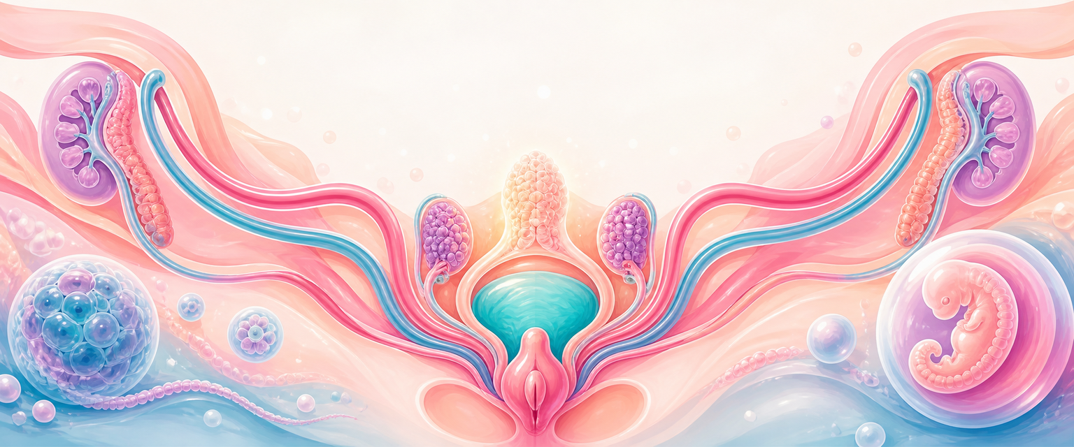
Urogenital and Reproductive Tract Development
Start with one sentence that the whole chapter hangs on: the urinary tract and the genital tract are built from the same strip of embryonic tissue, at the same time, by the same neighbours — so they fail together and you must always check both. Almost every clinical surprise in this field is a consequence of that single fact.
Because they share an origin, a malformed uterus often comes with a missing or misplaced kidney, and the link is developmental, not coincidental. A newborn with ambiguous genitalia cannot be understood without separately tracing gonadal differentiation, internal ducts, hormones and external genital patterning. A young woman with primary amenorrhoea and otherwise normal 46,XX puberty can have a completely absent uterus, because the ovaries and the uterus come from different embryonic structures that can succeed or fail independently.
The central map runs from a single tissue to the finished anatomy:
intermediate mesoderm -> urogenital ridge -> nephric system + gonadal ridge -> Wolffian and Mullerian ducts -> internal genital tract -> urogenital sinus and genital tubercle -> external genitalia
The key is to keep four layers separate:
| Layer | What develops | Clinical question |
|---|---|---|
| Chromosomal sex | XX, XY or sex-chromosome variation | What genetic programme is possible? |
| Gonadal sex | Ovary, testis, dysgenetic gonad, ovotestis | What hormones can the gonad produce? |
| Internal duct sex | Mullerian and/or Wolffian derivatives | Are uterus, tubes, cervix, upper vagina, epididymis or vas present? |
| External genital phenotype | Urethra, phallus/clitoris, labioscrotal folds, lower vagina | What did androgen exposure do at the target tissue? |
Most exam errors come from mixing these layers.
The practical clinical chain is:
karyotype/genes -> gonad -> hormones -> ducts -> external genitalia -> puberty/fertility/obstruction presentation
If the layers do not match, do not force them to match. Work layer by layer.
A second anchor that makes the whole story easier is timing. The events are not simultaneous; they happen in a fixed order across the embryonic and early fetal weeks, and an insult is only relevant if it lands during the window in which that structure is forming. A rough timeline worth carrying:
| Approximate gestational age | Event |
|---|---|
| ~4 weeks | Pronephros appears and regresses; primordial germ cells reach the genital ridge |
| ~5 weeks | Mesonephros and genital ridge present; first sign of the indifferent gonad; cloaca begins to be partitioned |
| ~5-6 weeks | Paramesonephric (Mullerian) ducts begin to form alongside the Wolffian ducts; ureteric bud sprouts |
| ~6-7 weeks | Bipotential ("indifferent") gonad fully assembled; SRY can now act |
| ~7 weeks | If SRY acts, the testis is first recognisable (Sertoli cells appear first) |
| ~9 weeks | Mullerian ducts reach the urogenital sinus; sex of external genitalia still not visible |
| ~10 weeks | Ovary first clearly identifiable; external genitalia start to differentiate by sex |
| ~12-16 weeks | Mesoderm around the fused ducts forms uterine and cervical muscle |
| ~16-18 weeks | Solid vaginal plate canalises into a lumen |
| ~20 weeks | Oogonia peak (~7 million) then decline by atresia |
You do not need to memorise the table to the week, but the order is examinable and clinically useful: the gonad commits before the ducts respond, and the ducts are laid down before the external genitalia take a recognisable shape.
Intermediate Mesoderm and the Urogenital Ridge
Now build from the tissue. The urinary and genital systems arise largely from intermediate mesoderm — the thin strip of mesoderm lying between the paraxial somites and the lateral plate. On each side of the midline, beneath the lining of the coelomic cavity, the intermediate mesoderm thickens into a paired urogenital ridge. Each urogenital ridge has two longitudinal territories side by side: a nephrogenic component (which becomes the urinary apparatus) and a gonadal (genital) ridge component on its medial aspect. This shared real estate is the embryological reason the two systems are inseparable in clinical practice.
Three kidney systems appear sequentially within the nephrogenic territory:
| System | Main point | Adult relevance |
|---|---|---|
| Pronephros | A few transient cervical tubules; appears first (~4 weeks) and quickly degenerates | No adult kidney contribution |
| Mesonephros | Temporary embryonic kidney in the thoracolumbar region; drained by the mesonephric (Wolffian) duct | Wolffian duct contribution in males; helper role in reproductive tract development |
| Metanephros | Definitive kidney, formed from ureteric bud plus metanephric mesenchyme | Adult kidney |
The mesonephric duct is not just a male duct. It is needed early as a guide and neighbour for other structures: the ureteric bud sprouts from it to make the kidney, and the developing paramesonephric (Mullerian) duct runs alongside it and uses it as a track to migrate caudally. So the mesonephric duct is the common scaffold for the whole urogenital field — which is exactly why a one-sided fault in this system tends to damage both the kidney and the genital tract on that side, and why urinary tract and genital tract anomalies travel together.
Why One Embryological Field Creates Many Presentations
The urogenital ridge contains kidney, duct and gonadal territories close together. A developmental disturbance can therefore appear as:
| Main presenting problem | Hidden linked system to check |
|---|---|
| Mullerian anomaly | renal tract |
| Renal agenesis/ectopic kidney | ipsilateral Mullerian/outflow anomaly if symptoms suggest |
| Ambiguous genitalia | adrenal/endocrine/electrolytes and internal ducts |
| Primary amenorrhoea | uterus, vagina, ovaries, hormones and karyotype |
| Cyclic pelvic pain in adolescent | obstructed uterus/vagina/hemivagina and renal association |
This is why "one ultrasound finding" often becomes a multi-system assessment.
Kidney and Ureter Development
The definitive kidney forms through interaction between:
- the ureteric bud, an outgrowth from the mesonephric duct;
- the metanephric mesenchyme, which forms nephron tissue.
The ureteric bud forms the collecting system: ureter, renal pelvis, calyces and collecting ducts. The metanephric mesenchyme forms nephrons: glomeruli, proximal tubules, loops of Henle and distal tubules. Development requires reciprocal induction. If the ureteric bud fails to form or reach the mesenchyme, the kidney may be absent. If branching is abnormal, collecting-system anomalies occur.
| Developmental failure | Possible anomaly | O&G relevance |
|---|---|---|
| Ureteric bud absent or fails to contact metanephric mesenchyme | Renal agenesis | Screen kidneys with Mullerian anomalies |
| Abnormal ureteric bud position | Duplex system, ectopic ureter | Continence, recurrent UTI, pelvic surgery risk |
| Failed ascent/rotation | Pelvic kidney, malrotation | Pelvic mass differential; operative risk |
| Abnormal fusion | Horseshoe kidney | Vascular and positional variation |
| Persistent cloacal/urogenital sinus abnormality | Complex lower tract anomaly | Paediatric gynae/urology referral |
Kidneys ascend from the pelvis to the abdomen as the embryo grows. They do not "climb" actively so much as change relative position. Blood supply changes during ascent; accessory renal vessels reflect vessels that persist.