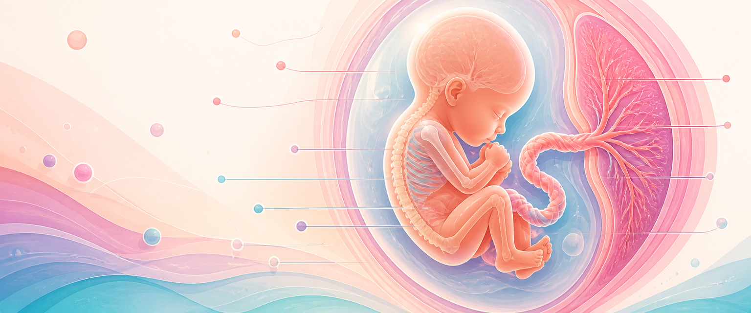
Anatomy of the Fetus
Start from one idea: the fetus is built around the fact that its oxygen comes from the placenta, not its own lungs. Everything that makes fetal anatomy different from adult anatomy flows from that single constraint. The lungs are fluid-filled and do no gas exchange, so blood is plumbed to bypass them. The liver gets first call on the richest blood, so a shunt diverts flow past it. The heart pumps in a way no adult heart does. The head is large and the skull is deliberately deformable because it has to be both grown fast and delivered through a bony pelvis. Hold that organising principle and the rest of fetal anatomy stops being a list and becomes a logic.
So fetal anatomy is developmental anatomy in motion, organised around placental life. The fetus is not a small adult: it has a large head, open cranial sutures, fluid-filled lungs, three temporary circulatory shunts, a placenta-dependent circulation, active growth plates, changing body proportions and organs whose form and function mature at different speeds. O&G uses this anatomy every day when interpreting ultrasound planes, counselling after an anomaly scan, assessing fetal growth, planning delivery, understanding labour mechanics and preparing for the neonatal transition.
The Primary candidate should be able to move between four views of any structure:
- developmental anatomy: how the structure formed;
- ultrasound anatomy: how the structure is recognised;
- obstetric anatomy: why it matters in pregnancy or labour;
- neonatal anatomy: what changes when placental support ends.
| Region | What to recognise | Why O&G cares |
|---|---|---|
| Head and CNS | Skull, sutures, ventricles, midline, posterior fossa | Anomaly scan, moulding, fetal lie and position |
| Face and neck | Orbits, profile, lips, palate, mandible, neck masses | Clefting, airway risk, chromosomal markers |
| Thorax and heart | Situs, four chambers, outflows, diaphragm, lungs | Congenital heart disease, lung hypoplasia, CDH |
| Abdomen | Stomach, bowel, liver, wall, cord insertion | Swallowing, obstruction, anterior wall defects |
| Renal tract and bladder | Kidneys, renal pelvis, bladder cycling, liquor | Urine production, obstruction, renal agenesis |
| Skeleton and movement | Spine, long bones, hands, feet, posture, tone | Skeletal dysplasia, neuromuscular disease, presentation |
The art is to avoid a list-only answer. A better answer says: this structure, this developmental job, this scan sign, this clinical implication.
The Fetal Circulation and the Three Shunts
Before looking at any organ in isolation, fix the circulation, because it is the feature that ties fetal anatomy together. In adult anatomy the right heart serves the lungs and the left heart serves the body, in series. In the fetus the lungs are fluid-filled and almost no blood needs to go through them, so the design is different: oxygenated blood arrives from the placenta, and three shunts steer it past the organs that do not yet need it.
Trace the path once and it stays learned:
- Oxygen-rich blood leaves the placenta in the single umbilical vein and enters the abdomen at the cord insertion.
- The first shunt, the ductus venosus, lets most of this blood bypass the liver sinusoids and pass straight into the inferior vena cava, so the richest blood is not diluted before it reaches the heart.
- That well-oxygenated stream enters the right atrium and is preferentially directed across the second shunt, the foramen ovale, into the left atrium, left ventricle and ascending aorta. This delivers the best-oxygenated blood to the coronary arteries, brain and upper body.
- Deoxygenated blood returning from the head via the superior vena cava enters the right atrium at a different angle, stays separate, and passes to the right ventricle and pulmonary artery. The two streams mix surprisingly little.
- Because the lungs are not for gas exchange yet, the third shunt, the ductus arteriosus, carries most right-ventricular output from the pulmonary artery directly into the descending aorta, bypassing the high-resistance pulmonary bed. Only a small fraction of the output actually perfuses the lungs.
- Blood then returns to the placenta for re-oxygenation through the paired umbilical arteries, which arise from the internal iliac arteries.
| Shunt | What it bypasses | Why it exists |
|---|---|---|
| Ductus venosus | Liver sinusoids | Keep placental blood richly oxygenated for the heart and brain |
| Foramen ovale | Right ventricle and lungs | Send the best blood left-sided, to coronaries, brain and upper body |
| Ductus arteriosus | Fluid-filled, high-resistance lungs | Divert right-heart output to the body and placenta, not the lungs |
Two further points separate the fetal heart from the adult heart and explain a lot of fetal physiology. First, the ventricles work in parallel, not in series: both sides feed the systemic circulation, so we speak of combined cardiac output rather than equating left ventricular output with cardiac output. The right ventricle does slightly more of the work than the left in the normal fetus. Second, this parallel arrangement gives the circulation real plasticity. When oxygen falls, the fetus can redistribute blood toward the brain, heart and adrenals and away from the periphery — the basis of the brain-sparing response that O&G monitors with Doppler later in pregnancy. The umbilical vein, the ductus venosus and the middle cerebral artery are exactly the vessels interrogated when assessing a growth-restricted fetus, which is why this anatomy is worth holding before anything else.
This is also where ultrasound anatomy and obstetric anatomy meet. A single umbilical artery instead of two is a real anatomical finding that should prompt a careful search for associated cardiac and renal anomalies. The ductus venosus is a named Doppler target, not an academic curiosity. And the whole logic of the outflow-tract cardiac views (described below) is to confirm that the great vessels — the very vessels that the shunts depend on — are connected correctly.
Fetal Head and Skull
The fetal head is large relative to the body, especially earlier in gestation. The skull vault contains sutures and fontanelles that allow growth and moulding. The clinically important landmarks are the anterior fontanelle, posterior fontanelle, sagittal suture, coronal sutures and lambdoid sutures.
It helps to name the regions of the vault, because labour terminology uses them. The occiput is the area behind the posterior fontanelle; the vertex is the area between the two fontanelles, over the parietal eminences; the bregma surrounds the anterior fontanelle; and the sinciput lies in front of it, subdividing into brow and face above and below the root of the nose. The vault bones form in membrane (allowing moulding), while the skull base forms in cartilage.
In labour, these landmarks allow assessment of position and flexion, and the degree of flexion decides which diameter must negotiate the pelvis. A well-flexed vertex presents the suboccipitobregmatic diameter, about 9.5 cm — the smallest anteroposterior diameter and the reason flexion matters so much. As the head deflexes, larger diameters present: the occipitofrontal diameter (about 11.5 cm) with a deflexed vertex, and the mentovertical diameter (about 13 cm) with a brow presentation, which is the largest and least favourable. A face presentation again presents a relatively small diameter (submentobregmatic, of the order of 9.5 cm) once fully extended. Deflexion therefore increases the presenting diameter and can cause slower progress or obstruction. Moulding allows the parietal bones to slide under each other and the frontal and occipital bones to slide under the parietals, which can reduce the presenting dimension by roughly 1 to 1.5 cm; this is physiological in normal labour, but excessive moulding can signal obstruction or prolonged pressure.