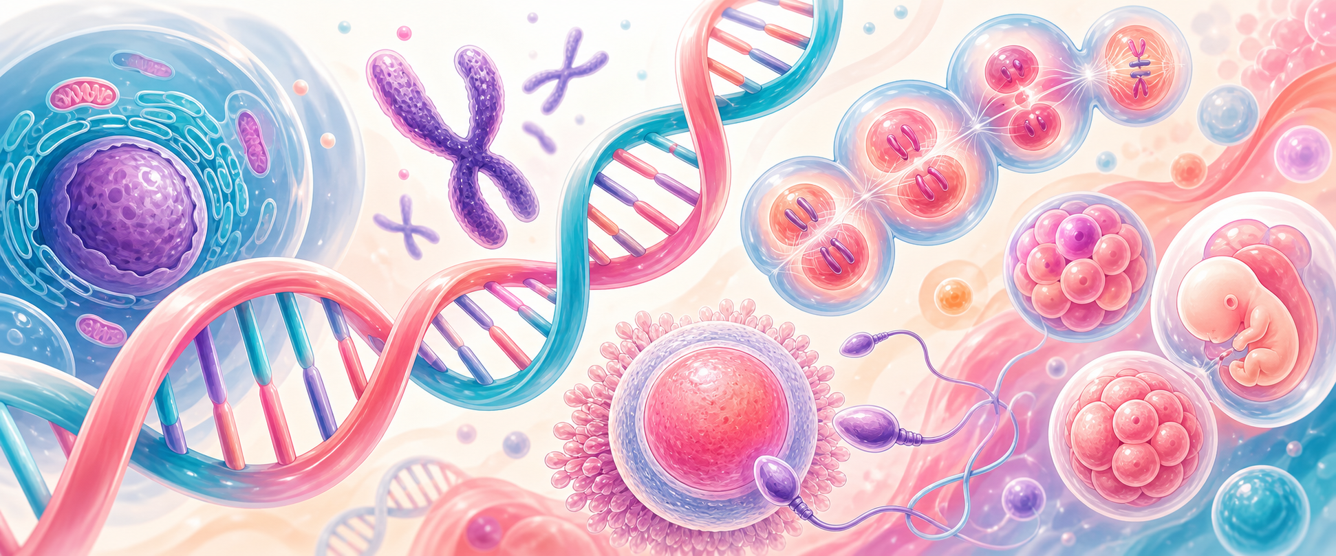
Cell Biology for Embryology
Start with one sentence the rest of the chapter hangs from: every cell carries a complete copy of the genome, and development is the controlled copying, distribution and reading of that genome over time. A new human being is not built by adding new information at each step; it is built by faithfully duplicating the same DNA, dividing it correctly between cells, and switching different parts of it on in different places. Almost every O&G genetics problem you will meet is a failure of one of those three jobs — copy it, distribute it, or read it.
Embryology therefore begins before an embryo is visible. It begins with DNA being copied, chromosomes being separated, gametes being formed, and two haploid genomes being combined. If this foundation is weak, later topics such as aneuploidy, miscarriage, congenital anomaly, molar pregnancy, infertility, cancer and prenatal screening feel like disconnected facts. They are not. They are consequences of how cells store, copy and distribute genetic information.
We will build in the order a cell itself uses. First, how information is stored and read (DNA structure, then the central dogma). Then how it is packaged and counted (chromosomes). Then how it is copied and divided (the cell cycle, mitosis). Then how it is halved to make gametes (meiosis and gametogenesis). Then how two halves are recombined (fertilisation). Only after that do we apply the mechanism to the clinical layer — abnormalities, inheritance patterns, genetic tests, imprinting and the O&G situations where this all becomes a counselling conversation.
For the Primary exam, the essential chain is:
- DNA stores information in base sequence;
- that sequence is read out as RNA and protein (the central dogma);
- cells duplicate DNA during the cell cycle;
- mitosis distributes identical chromosomes to daughter cells;
- meiosis halves chromosome number to make gametes;
- recombination creates variation but also creates segregation risk;
- fertilisation restores diploidy;
- early development depends on repeated mitotic divisions and lineage decisions.
| Process | What it does | O&G consequence when it goes wrong |
|---|---|---|
| DNA replication | Copies the genome before division | Replication error, mutation, failed early embryo development |
| Mitosis | Preserves chromosome number in daughter cells | Mosaicism, abnormal growth, neoplasia if control fails |
| Meiosis I | Separates homologous chromosomes | Non-disjunction, aneuploid gamete |
| Meiosis II | Separates sister chromatids | Aneuploid gamete if separation fails |
| Fertilisation | Restores diploidy and activates development | Triploidy, molar pregnancy, failed fertilisation |
DNA Structure and Base Pairing
DNA is a double-stranded polymer built from nucleotides. Each nucleotide contains a sugar (deoxyribose), a phosphate group and a nitrogenous base. The bases are adenine, thymine, cytosine and guanine. Adenine pairs with thymine. Cytosine pairs with guanine. This complementary pairing is held by hydrogen bonds and is the single most important property of DNA, because it means each strand carries enough information to rebuild its partner. That is what allows DNA to be copied accurately, generation after generation and cell after cell.
The two strands run in opposite directions — they are antiparallel — and twist into a double helix, with the bases stacked on the inside. The sequence of bases encodes genes and regulatory regions. A gene is not simply a "trait"; it is a DNA sequence that can be transcribed into RNA and, for many genes, translated into protein. Regulatory DNA helps decide when, where and how strongly genes are expressed. This matters in embryology because development is not achieved by changing the genome in every cell; it is achieved by controlling which genes are switched on in which cells at which time.
Copying is semiconservative: the two strands separate, and each acts as a template for a new complementary strand, so each daughter molecule keeps one old strand and gains one new one. Replication happens during one specific window of the cell's life (the S phase, described below), not during chromosome separation — a distinction that examiners test and that explains why a replication error and a segregation error are different categories of fault. If the copying machinery makes a mistake that escapes repair, the result is a mutation (a permanent sequence change). If the dividing machinery mis-distributes whole chromosomes, the result is aneuploidy (a wrong chromosome count). Both can damage a pregnancy, but they happen at different steps and by different mechanisms.
In O&G, DNA structure becomes clinically relevant in several ways:
- single-gene variants can cause inherited disease;
- chromosomal aneuploidy can cause miscarriage or fetal syndromes;
- abnormal imprinting and parental genomic contribution can contribute to molar pregnancy;
- DNA damage and failed repair can contribute to neoplasia;
- fetal DNA fragments in maternal plasma allow screening for common aneuploidies.
The key exam distinction is that DNA sequence, chromosome number and gene expression are different layers. A fetus may have a normal sequence but abnormal chromosome number. A tissue may have the same DNA as another tissue but different gene expression. A cancer may arise from acquired genetic and epigenetic changes rather than inherited disease.
The Central Dogma: How the Sequence Is Read
Before chromosomes and division make sense, it helps to know what the sequence is actually for. The classic summary is the central dogma: DNA → RNA → protein. DNA is the stored archive; it stays in the nucleus and is rarely used directly. To use a gene, the cell first transcribes the relevant stretch of DNA into a working copy of messenger RNA (mRNA), then translates that mRNA into a chain of amino acids that folds into a protein. Proteins are the workers — enzymes, receptors, structural fibres, hormones — so almost everything a cell does is the downstream consequence of which genes get read.
A few details earn their place because they explain disease:
- The genetic code is read in three-base units called codons; each codon specifies one amino acid, and the code is redundant (several codons can mean the same amino acid), which is why the third base of a codon is often not critical.
- A protein-coding gene is split into exons (the coding pieces) and introns (intervening sequences). The first RNA copy contains both; splicing removes the introns and joins the exons into mature mRNA. A variant that disrupts a splice site can therefore disable a gene without touching a single codon.
- Translation happens on ribosomes, where transfer RNA (tRNA) molecules bring the correct amino acid for each codon.
- Not all RNA codes for protein. tRNA and ribosomal RNA build the machinery itself, and a large family of non-coding RNAs help regulate which genes are expressed.
This is why "a variant in the DNA" is not one thing. A change can alter a codon (and the protein), create a premature stop, shift the reading frame, disrupt splicing, change gene dosage, or alter only the regulatory DNA that decides when and where a gene is read. We return to those categories once chromosomes are in place — but the principle to carry forward is that the genome is information that has to be copied, distributed and read, and a fault at any of those steps can present in O&G.
Chromosome Language
Human somatic cells are usually diploid: 46 chromosomes arranged as 23 pairs. Gametes are haploid: 23 chromosomes. The autosomes are chromosomes 1 to 22. The sex chromosomes are X and Y. The terms matter because many O&G conditions are chromosome-number conditions rather than single-gene conditions.
| Term | Meaning | O&G example |
|---|---|---|
| Euploid | Whole sets of chromosomes present | Normal 46,XX or 46,XY conceptus |
| Aneuploid | One or more chromosomes gained or lost | Trisomy 21, monosomy X |
| Polyploid | Extra whole chromosome set | Triploidy, partial molar patterns |
| Mosaicism | Two or more cell lines from one zygote | Placental mosaicism affecting NIPT or CVS interpretation |
| Chimera | Cell lines from more than one zygote | Rare; not the usual explanation for mosaic results |
Aneuploidy is a leading cause of early pregnancy loss. The embryo often arrests because gene dosage is incompatible with normal development. Some aneuploidies can survive to later pregnancy or birth because the gene-dosage disturbance is compatible with survival, but not normal development. This explains why miscarriage tissue, NIPT, CVS and amniocentesis all sit on the same biological foundation: chromosome counting and cell lineage.
Mosaicism requires a timing concept. If an error happens in meiosis before fertilisation, every cell of the conceptus starts with the abnormal chromosome number. If an error happens during an early mitotic division after fertilisation, the embryo may contain more than one cell line. If the abnormal line is mainly placental, the fetus may be different from the placenta. That is why placental tests can require careful confirmation.
Genome Organisation and Gene Expression
The genome is not loose DNA floating in the nucleus. DNA is packaged with histone proteins into chromatin, condensed into chromosomes when cells divide, and controlled by regulatory regions that decide when genes are used. This packaging matters because development depends on switching genes on and off in the right cell at the right time.
| Genome layer | Meaning | O&G relevance |
|---|---|---|
| Chromosome | Long DNA molecule packaged with proteins | Aneuploidy, translocation, deletion and duplication |
| Centromere | Constricted region needed for spindle attachment | Mis-segregation can cause aneuploidy |
| Telomere | Protective chromosome end | Genome stability and cellular ageing concepts |
| Chromatin | DNA plus histone and associated proteins | Open chromatin is more transcriptionally active |
| Gene | DNA region that produces a functional RNA or protein product | Single-gene disorders and developmental patterning |
| Regulatory DNA | Promoters, enhancers and silencers controlling expression | Same DNA can behave differently in different tissues |
Building on the central dogma above, a protein-coding gene sits in a controlled environment: a promoter (which sets basal activity, usually just upstream of the gene) and enhancers and silencers (which tune how strongly and in which tissue the gene is read). This regulatory layer is why the same genome can run a heart cell, a neuron and a trophoblast — the difference is which genes are switched on, not which genes are present. It is also why a disease-causing variant does not have to sit in the coding sequence at all. A variant can change protein sequence, create a premature stop, shift the reading frame, disrupt splicing, alter gene dosage, or disturb the regulatory DNA that controls when and where a gene is expressed.