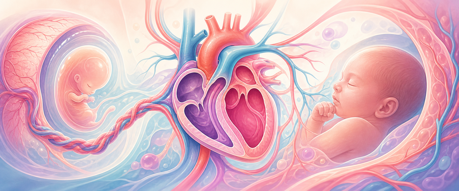
Cardiovascular Development and Fetal Circulation
Start with the single fact that everything else hangs on: the embryo outgrows diffusion before it has a finished heart. A ball of cells can absorb oxygen and nutrients straight across its surface, but once the embryo is more than a couple of millimetres thick, the cells in the middle starve. There is no time to wait for a tidy four-chamber heart. So the cardiovascular system becomes the first organ system that has to work while it is still being built — it must pump before it is finished.
That single constraint explains the whole rest of this chapter. Because building and pumping happen at the same time, the heart is assembled as a moving target: blood vessels form (angiogenesis), a tube forms, the tube bends (looping), chambers balloon, walls grow (septation), the outflow twists and divides, valves carve out, and the great vessels remodel — all while the tube is already contracting and while the placenta, not the lung, is doing gas exchange. Build a complicated machine while it is running and you will accept compromises; in the fetus those compromises are the fetal shunts, deliberate holes and bypasses that are normal before birth and must close after it.
For O&G this chapter does four jobs, and they stack on top of one another:
- explain why congenital heart defects cluster around a few developmental events — looping, septation, the outflow tract and the neural crest;
- explain why the fetal heart depends on the placenta, not the lungs, and beats as two pumps in parallel rather than in series;
- explain why fetal hypoxia changes heart-rate patterns and blood-flow distribution before it causes terminal acidosis — the basis of all fetal monitoring;
- prepare you for fetal anomaly scanning, fetal heart-rate interpretation, neonatal transition and adult congenital heart disease in pregnancy.
The single mental model to carry through the chapter is the assembly line, each step depending only on the one before it:
cardiogenic mesoderm → paired heart tubes → single heart tube → looping → chamber ballooning → septation → outflow rotation → valve formation → fetal shunts → birth transition
Each step is a place where development can fail, and each failure produces a recognisable lesion. Learn the order and the lesions almost predict themselves.
A Timeline You Can Reason from
Before the mechanism, fix the calendar, because timing is the single most useful clinical tool in this chapter. The whole heart is built in a remarkably short window.
| Time after fertilisation | Cardiovascular event | Why O&G cares |
|---|---|---|
| Week 3 | Cardiogenic field and blood islands appear | Diffusion is failing; a circulation becomes essential |
| Day 20-22 | Paired endocardial tubes form and begin to fuse | Tube-formation defects are usually severe |
| Day 22-23 | Primitive heart tube begins to beat | Cardiac activity is detectable on ultrasound from around day 32 |
| Week 4 | Cardiac looping | Laterality and chamber-position defects arise here |
| Weeks 4-5 | Chamber ballooning and endocardial cushions | AV-septal and valve defects link to cushion biology |
| Weeks 5-7 | Atrial, ventricular and outflow septation | The main window for major congenital heart disease |
| Weeks 7-8 | Semilunar valves and great-vessel remodelling | Outflow and arch anomalies become recognisable |
| Fetal period | Growth, maturation, shunt physiology, reserve | Functional surveillance dominates |
| Birth | Pulmonary resistance falls; shunts close | Neonatal transition and cyanosis are explained here |
The structural heart is essentially complete between the fourth and seventh weeks after fertilisation. Hold one conversion in your head, because examiners and clinics use different clocks: embryological age is about two weeks less than obstetric (gestational) age, which is dated from the last menstrual period. A woman who is "six weeks pregnant" by LMP is about four weeks past fertilisation — exactly when the tube is looping and septation is starting. This is why a teratogenic or metabolic insult around the time the pregnancy is first recognised lands squarely on cardiac organogenesis, and why preconception care, not booking care, is where structural prevention happens.
The Cardiogenic Field and the Heart Tube
Now build the machine, beginning with the raw material. After gastrulation, cardiac progenitor cells migrate out and settle as mesoderm in the cranial part of the embryonic disc. They organise into two related populations rather than one. The primary (first) heart field contributes much of the early left ventricle and the atria. The second heart field adds cells later to the outflow tract, the right ventricle and parts of the inflow. The reason to know this is purely clinical: the heart is not made by one static tube that simply folds up. Cells are added as the heart elongates, so faults in adding or patterning the second-heart-field contribution show up specifically as outflow-tract and right-sided defects — a recurring theme below.
The earliest heart is a pair of endocardial tubes lying in the splanchnic (visceral) mesoderm on either side of the midline. Two folding movements bring them together. Lateral folding sweeps the paired tubes toward the midline, where they fuse into a single primitive heart tube. Cranial-caudal (head-to-tail) folding then carries the cardiogenic region down from its initially cranial position into the future thorax, and bends the tube so its venous (inflow) end and arterial (outflow) end start to come close. The tube is built of an outer myocardium and an inner endocardium, separated by gelatinous cardiac jelly that will later contribute to cushions and valves.
The straight tube has named regions, each of which becomes a defined adult structure. You do not memorise this as a list; you use it to locate lesions along the inflow-to-outflow axis.
| Primitive region (inflow → outflow) | Later contribution | Clinical bridge |
|---|---|---|
| Sinus venosus | Smooth-walled right atrium and the venous inflow | Sinus-venosus ASD, anomalous venous return |
| Primitive atrium | Trabeculated parts of both atria and the appendages | Atrial septation and appendage anatomy |
| Primitive ventricle | Trabeculated left ventricle | Ventricular septal anatomy |
| Bulbus cordis | Smooth right-ventricular outflow and the conotruncal region | Tetralogy, transposition, double-outlet patterns |
| Truncus arteriosus | Roots of the aorta and pulmonary trunk | Persistent truncus, conotruncal defects |
The tube is initially a straight pump with inflow at one end and outflow at the other. To turn that linear arrangement into a heart with correctly placed right and left structures, it has to bend. That bending is the next step.
Looping and Laterality
By day 23 the tube is contracting; in week 4 it loops. Normal looping bends the tube to the right (a "D-loop"), so that the future right and left ventricles and the outflow tract end up in their correct spatial relationships. Looping is the first visible sign of left-right asymmetry in the whole embryo, and it is governed by the same body-wide patterning machinery that places the stomach, spleen, liver and lungs. That shared control is why cardiac laterality defects so often travel with abnormal positioning of the abdominal organs.
| Looping / laterality problem | Mechanistic idea | O&G relevance |
|---|---|---|
| Dextrocardia | Heart apex points to the right | May be isolated or part of a situs abnormality |
| Situs inversus | Complete mirror-image organ arrangement | Can coexist with ciliary (cilia-driven) disorders |
| Heterotaxy | Disordered, incomplete left-right patterning | High risk of complex congenital heart disease |
| Malpositioned outflow | Abnormal ventricle-to-great-artery relationship | Transposition-type physiology |
Looping teaches an examinable principle that recurs at the anomaly scan: cardiac anatomy is three-dimensional, and the relationships are set very early. A normal-looking four-chamber view does not exclude an outflow-tract lesion, because the outflow relationships are established by looping and outflow rotation — events the four-chamber view does not show. This is why outflow-tract views are a required part of fetal cardiac assessment, not an optional extra.
Septation: Atria, Ventricles and the Endocardial Cushions
Once the tube has looped, the single channel must be partitioned into right and left sides. But here is the fetal twist: it must be partitioned while preserving deliberate communications so that the parallel fetal circulation can still work. Septation is therefore not crude wall-building; it is controlled partitioning that leaves designed gaps.
Three regions septate at roughly the same time: the atria, the ventricles, and the atrioventricular (AV) canal between them. The endocardial cushions — swellings of cardiac jelly on the dorsal and ventral walls of the AV canal — are the hinge of the whole process. They grow inward and fuse, dividing the common AV canal into right and left, and they also contribute to the lower part of the atrial septum, the membranous part of the ventricular septum, and the AV valves. Because one tissue does so many jobs, a single cushion defect can produce a combined atrial-ventricular-valve abnormality — which is exactly what an AV septal defect is.