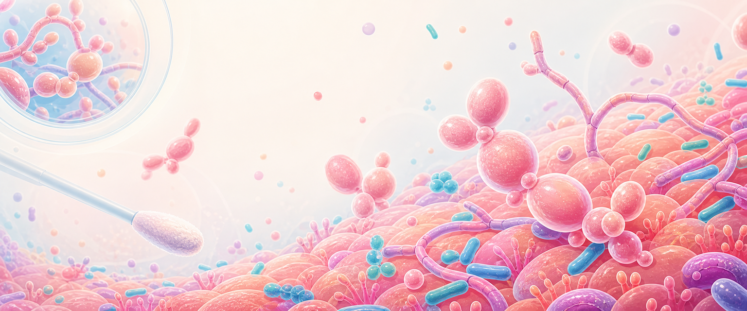
Yeast Infections
Start with one idea and let everything else hang off it: a yeast is a fungus, and a fungus is not a bacterium. That single distinction predicts almost everything you need for the exam — which drugs work, which do not, why the vaginal pH stays normal, why a positive swab is not the same as disease, and why repeated antibacterial courses can cause thrush. If you anchor on "different kind of cell, different rules", the rest of this chapter is just unpacking the consequences.
In O&G the syndrome you will be asked about is vulvovaginal candidiasis (VVC), almost always caused by Candida albicans. But before any of the clinical detail makes sense, you need the biology underneath it. So this chapter builds in deliberate steps: first what a fungus is, then how Candida lives on the vaginal mucosa, then how a harmless coloniser tips over into inflammatory disease, then how you confirm it, distinguish it from its many mimics, and reason about treatment — all from mechanism rather than memorised regimens.
What a Fungus Is (and Why That Decides the Treatment)
A fungus is a eukaryote: it has a true nucleus and organelles, like our own cells, and unlike bacteria. That family resemblance is the whole problem of antifungal therapy — anything you use to kill the fungus risks harming the host, because the two cells are built on the same plan. This is the principle of selective toxicity, and it is the thread that runs through every antimicrobial answer: you have to find a structure or pathway the pathogen owns and the host does not.
So where do fungi differ from us? In two places that matter clinically.
| Fungal structure | What it is | Drug consequence |
|---|---|---|
| Cell wall | A rigid outer wall of chitin plus the polysaccharides glucan and mannan — not the peptidoglycan of bacteria, and absent from human cells altogether | A wall humans lack, so wall-synthesis inhibitors (echinocandins block glucan synthesis) are highly fungus-selective |
| Cell membrane | A lipid bilayer whose key sterol is ergosterol, where human membranes use cholesterol | The single most exploited antifungal target: azoles block ergosterol synthesis; polyenes bind ergosterol directly |
Notice what is not on that list. Fungi have no peptidoglycan, so beta-lactams (penicillins, cephalosporins) do nothing to Candida — they target a bacterial wall enzyme that fungi do not possess. Fungi have 80S ribosomes like ours, not the 70S ribosomes of bacteria, so antibacterial protein-synthesis drugs (aminoglycosides, macrolides, tetracyclines) also miss them. This is why you cannot treat thrush with an antibiotic, and why the reflex "discharge, so give an antibiotic" is a category error when the cause is yeast.
A short, memorable version: antibacterials attack walls and ribosomes that fungi do not share; antifungals attack the sterol membrane and chitin/glucan wall that human cells do not share. Same logic, opposite target.
How Fungi Grow and Reproduce
Fungi are generally larger than bacteria and many are multicellular. Most are aerobic, though many yeasts can ferment sugars to alcohol anaerobically — a clue to why a glycogen-rich, oestrogenised vagina is such good habitat. Most human pathogens grow over a wide temperature range; the species that cause invasive disease, including C. albicans, grow best at 37 °C, i.e. at body temperature. They reproduce mainly by asexual spores; C. albicans in particular forms tough chlamydospores that help it survive hostile conditions. In the laboratory, Candida and most pathogenic fungi grow readily on Sabouraud dextrose agar (and will grow on blood agar too) — which is exactly why a culture being positive is unremarkable and never, on its own, proof of disease.
The Four Shapes of Fungus
Pathogenic fungi sort into four morphological groups. You do not need encyclopaedic mycology for Primary O&G, but you do need to place Candida correctly and know why its shape is diagnostically useful.
| Group | Basic morphology | Example | O&G relevance |
|---|---|---|---|
| Moulds (filamentous) | Long branching hyphae weaving a mycelium; powdery spore-bearing colonies | Dermatophytes (ringworm); Aspergillus, Mucor in the immunocompromised | Skin/nail and opportunistic systemic disease — not routine vaginitis |
| True yeasts | Round/oval single cells reproducing by budding | Cryptococcus neoformans (large polysaccharide capsule) | Cryptococcal meningitis in advanced HIV — a reason WLWH are a special host group |
| Yeast-like fungi | Budding cells that also form branching pseudohyphae (and true hyphae) | Candida, chiefly C. albicans | The genus that causes VVC — the core of this objective |
| Dimorphic fungi | Yeast form in the body/at 37 °C, mould form in the environment/at 22 °C | Histoplasma capsulatum; Pneumocystis jirovecii (PJP) sits near this group | Specialist systemic mycology; PJP matters as an HIV-defining pneumonia |
Candida is therefore a yeast-like fungus: a budding yeast that can switch into elongated filaments. That switch is the single most important piece of Candida biology for the clinician, so it gets its own section.
Candida Morphology Is a Behaviour Report
C. albicans is polymorphic — it can live as round budding yeast, as pseudohyphae, or as true hyphae, and it shifts between them in response to its environment. This is not laboratory trivia. The shape the organism adopts tells you what it is doing:
- Budding yeast is the quiet, colonising state. It can sit on mucosa indefinitely without causing trouble.
- Pseudohyphae and hyphae are the invasive, tissue-engaging state. Filamentation drives adherence to epithelium, penetration between cells, and the inflammatory signalling that produces symptoms.
So when microscopy of a symptomatic patient shows pseudohyphae or hyphae, you are seeing the organism in its disease-causing form — far more convincing than a bare report of "Candida cultured". A useful confirmatory laboratory trick: C. albicans uniquely sprouts germ tubes when incubated in serum, which distinguishes it from most other Candida species. And C. albicans is acid-tolerant — it thrives at the normal acidic vaginal pH, which is why, unlike bacterial vaginosis (BV) and trichomoniasis, candidiasis classically occurs with a normal vaginal pH (≤ 4.5). That is one of the most useful bedside discriminators in any discharge question.
| Microscopy finding | Basic-science meaning | Clinical caution |
|---|---|---|
| Budding yeasts only | Colonising/quiescent state | May be colonisation, especially if no symptoms or signs |
| Pseudohyphae or hyphae | Invasive, tissue-engaging state | Supports symptomatic VVC when itch, erythema and normal pH align |
| No hyphae but symptoms persist | Some non-albicans species (e.g. C. glabrata) seldom filament | Speciate by culture rather than repeating blind azoles |
| Heavy growth on Sabouraud/blood agar | Candida grows readily | Growth alone never proves disease |